Research Group Computer Assisted Medicine (CaMed), Reutlingen University, 72762, Reutlingen, Germany.
Faculty of Electronic Engineering (FEE), Menoufia University, Menouf, 32952, Egypt.
Int J Comput Assist Radiol Surg. 2020 Jun;15(6):909-920. doi: 10.1007/s11548-020-02186-z. Epub 2020 May 5.
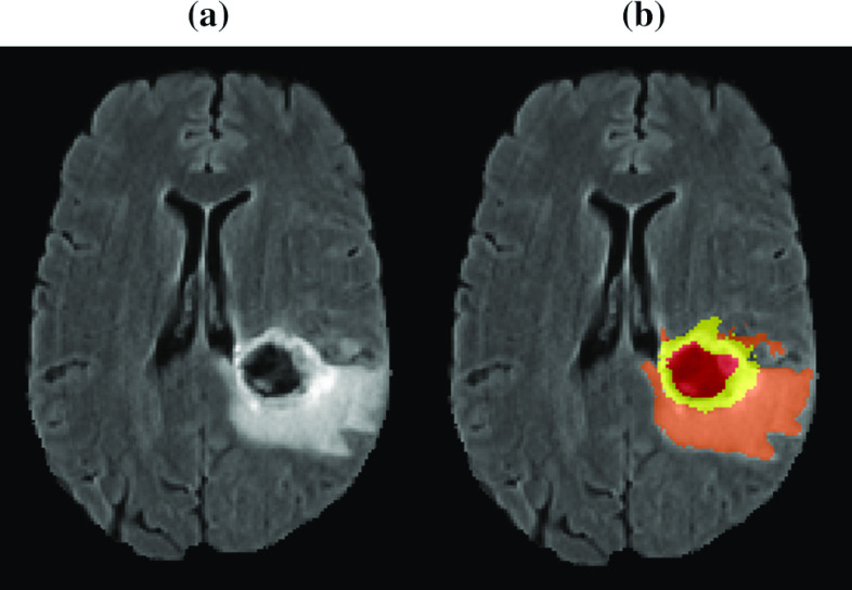
Gliomas are the most common and aggressive type of brain tumors due to their infiltrative nature and rapid progression. The process of distinguishing tumor boundaries from healthy cells is still a challenging task in the clinical routine. Fluid-attenuated inversion recovery (FLAIR) MRI modality can provide the physician with information about tumor infiltration. Therefore, this paper proposes a new generic deep learning architecture, namely DeepSeg, for fully automated detection and segmentation of the brain lesion using FLAIR MRI data.
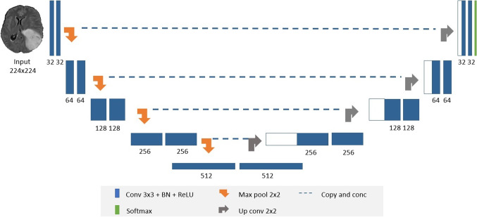
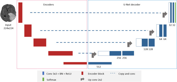
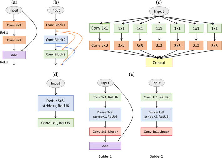
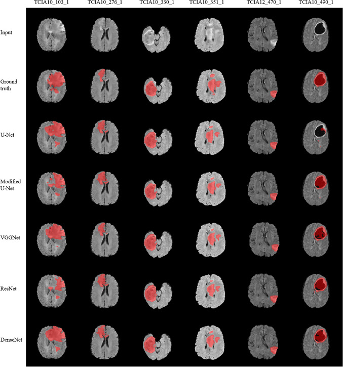
The developed DeepSeg is a modular decoupling framework. It consists of two connected core parts based on an encoding and decoding relationship. The encoder part is a convolutional neural network (CNN) responsible for spatial information extraction. The resulting semantic map is inserted into the decoder part to get the full-resolution probability map. Based on modified U-Net architecture, different CNN models such as residual neural network (ResNet), dense convolutional network (DenseNet), and NASNet have been utilized in this study.
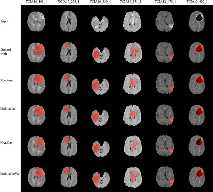
The proposed deep learning architectures have been successfully tested and evaluated on-line based on MRI datasets of brain tumor segmentation (BraTS 2019) challenge, including s336 cases as training data and 125 cases for validation data. The dice and Hausdorff distance scores of obtained segmentation results are about 0.81 to 0.84 and 9.8 to 19.7 correspondingly.
This study showed successful feasibility and comparative performance of applying different deep learning models in a new DeepSeg framework for automated brain tumor segmentation in FLAIR MR images. The proposed DeepSeg is open source and freely available at https://github.com/razeineldin/DeepSeg/.
由于胶质瘤具有浸润性和快速进展的特点,因此是最常见和最具侵袭性的脑肿瘤类型。区分肿瘤边界与健康细胞的过程在临床常规中仍然是一项具有挑战性的任务。液体衰减反转恢复(FLAIR)MRI 方式可以为医生提供有关肿瘤浸润的信息。因此,本文提出了一种新的通用深度学习架构,即 DeepSeg,用于使用 FLAIR MRI 数据自动检测和分割脑病变。
所开发的 DeepSeg 是一个模块化解耦框架。它由两个基于编码和解码关系的连接核心部分组成。编码部分是一个负责空间信息提取的卷积神经网络(CNN)。生成的语义图插入到解码器部分以获得全分辨率概率图。基于修改后的 U-Net 架构,本研究中使用了不同的 CNN 模型,如残差神经网络(ResNet)、密集卷积网络(DenseNet)和 NASNet。
所提出的深度学习架构已成功在线测试和评估,基于脑肿瘤分割(BraTS 2019)挑战赛的 MRI 数据集,包括 336 个病例作为训练数据和 125 个病例作为验证数据。获得的分割结果的骰子和 Hausdorff 距离得分分别约为 0.81 至 0.84 和 9.8 至 19.7。
本研究表明,在新的 DeepSeg 框架中应用不同的深度学习模型进行自动脑肿瘤分割 FLAIR MRI 图像具有成功的可行性和比较性能。所提出的 DeepSeg 是开源的,并可在 https://github.com/razeineldin/DeepSeg/ 上免费获得。