Lidenge Salum J, Tso For Yue, Ngalamika Owen, Kolape Jaydeep, Ngowi John R, Mwaiselage Julius, Wood Charles, West John T
Nebraska Center for Virology, Lincoln, NE, USA.
School of Biological Sciences, University of Nebraska-Lincoln, Lincoln, NE, USA.
Oncotarget. 2020 Apr 28;11(17):1556-1572. doi: 10.18632/oncotarget.27569.
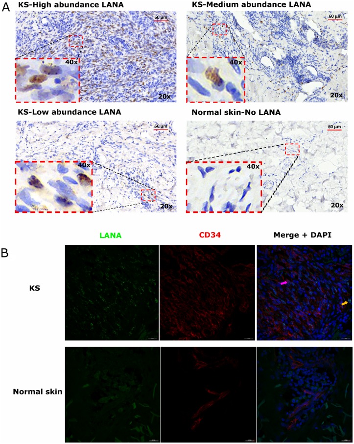
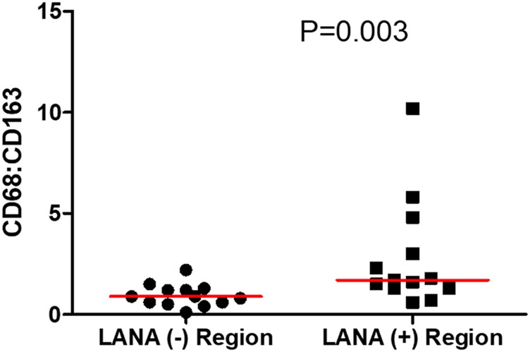
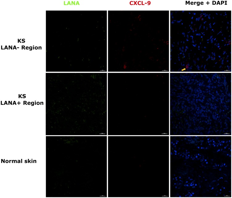
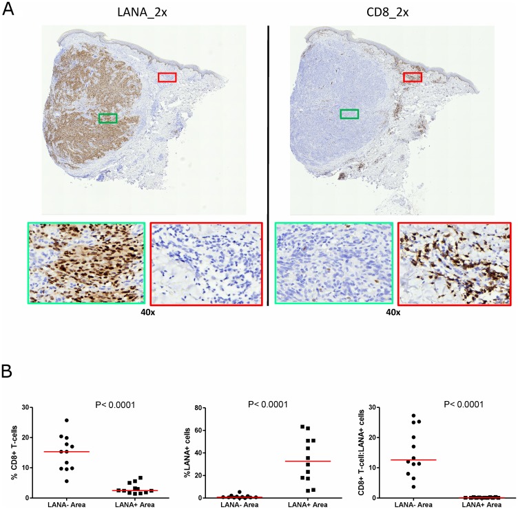
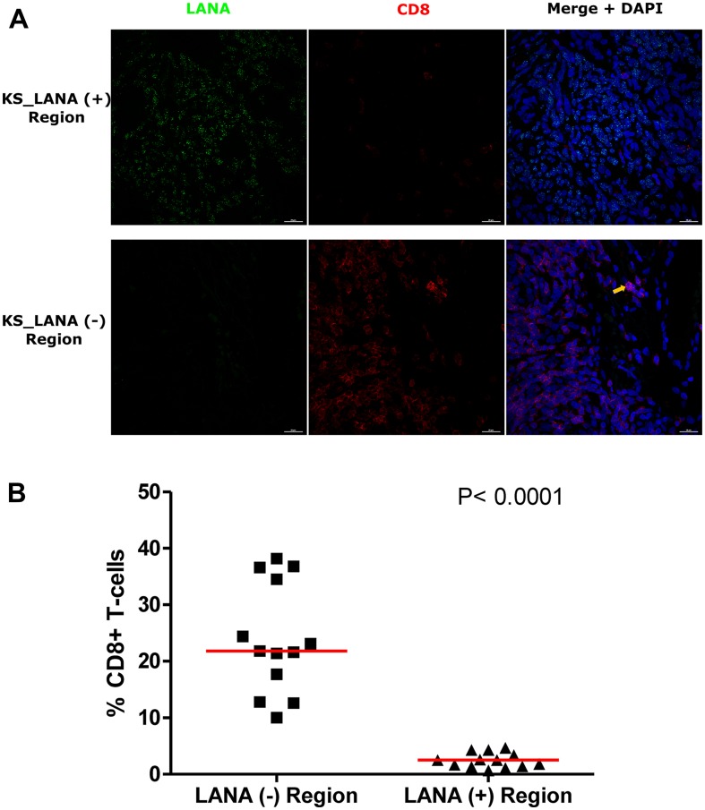
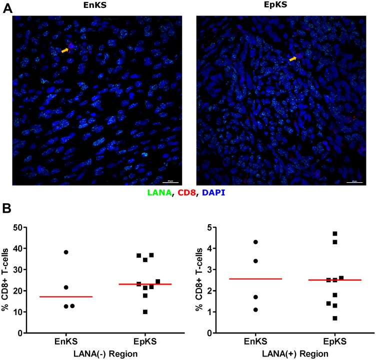
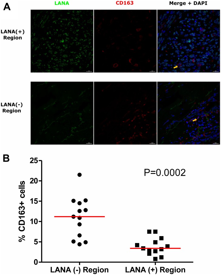
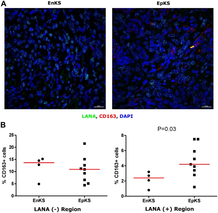
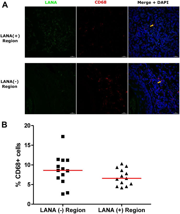
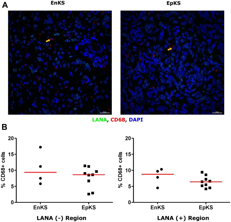
Despite the close association between Kaposi's sarcoma (KS) and immune dysfunction, it remains unclear whether tumor infiltrating immune cells (TIIC), by their absence, presence, or dysfunction, are mechanistically correlated with KS pathogenesis. Therefore, their potential capacity to serve as prognostic biomarkers of KS disease progression or control is unclear. Because epidemic-KS (EpKS) occurs with HIV-1 co-infection, it is particularly important to compare TIIC between EpKS and HIV-negative African endemic-KS (EnKS) to dissect the roles of HIV-1 and Kaposi Sarcoma-associated herpesvirus (KSHV) in KS pathogenesis. This cross-sectional study of 13 advanced KS (4 EnKS, 9 EpKS) patients and 3 healthy controls utilized single-color immunohistochemistry and dual-color immunofluorescence assays to characterize and quantify KSHV infected cells in relation to various TIIC in KS biopsies. Analysis of variance (ANOVA) and Mann-Whitney tests were used to assess differences between groups where -values < 0.05 were considered significant. The abundance of KSHV infected cells was heterogeneous in KS biopsies. Despite the presence of T-cell chemoattractant chemokine CxCL-9 in biopsies, CD8 T-cells were sparsely distributed in regions with evident KSHV infected cells but were readily detectable in regions devoid of KSHV infected cells ( < 0.0001). CD68 (M1) macrophages were evenly and diffusely distributed in KS biopsies, whereas, the majority of CD163 (M2) macrophages were localized in regions devoid of KSHV infected cells ( < 0.0001). Overall, the poor immune cell infiltration or co-localization in KS biopsies independent of HIV-1 co-infection suggests a fundamental tumor immune evasion mechanism that warrants further investigation.
尽管卡波西肉瘤(KS)与免疫功能障碍密切相关,但肿瘤浸润免疫细胞(TIIC)的缺失、存在或功能障碍是否在机制上与KS发病相关仍不清楚。因此,它们作为KS疾病进展或控制的预后生物标志物的潜在能力尚不清楚。由于流行性KS(EpKS)与HIV-1合并感染有关,比较EpKS和HIV阴性的非洲地方性KS(EnKS)之间的TIIC,以剖析HIV-1和卡波西肉瘤相关疱疹病毒(KSHV)在KS发病机制中的作用尤为重要。这项对13例晚期KS(4例EnKS,9例EpKS)患者和3名健康对照的横断面研究,利用单色免疫组织化学和双色免疫荧光分析来表征和量化KS活检中与各种TIIC相关的KSHV感染细胞。方差分析(ANOVA)和曼-惠特尼检验用于评估组间差异,其中P值<0.05被认为具有统计学意义。KS活检中KSHV感染细胞的丰度是异质性的。尽管活检中存在T细胞趋化因子趋化因子CxCL-9,但CD8 T细胞在有明显KSHV感染细胞的区域分布稀疏,而在没有KSHV感染细胞的区域很容易检测到(P<0.0001)。CD68(M1)巨噬细胞在KS活检中均匀且弥漫分布,而大多数CD163(M2)巨噬细胞位于没有KSHV感染细胞的区域(P<0.0001)。总体而言,KS活检中免疫细胞浸润不良或共定位与HIV-1合并感染无关,提示一种基本的肿瘤免疫逃逸机制值得进一步研究。