• 文献检索
  • 文档翻译
  • 深度研究
  • 学术资讯
  • Suppr Zotero 插件Zotero 插件
  • 邀请有礼
  • 套餐&价格
  • 历史记录
应用&插件
Suppr Zotero 插件Zotero 插件浏览器插件Mac 客户端Windows 客户端微信小程序
定价
高级版会员购买积分包购买API积分包
服务
文献检索文档翻译深度研究API 文档MCP 服务
关于我们
关于 Suppr公司介绍联系我们用户协议隐私条款
关注我们

Suppr 超能文献

核心技术专利:CN118964589B侵权必究
粤ICP备2023148730 号-1Suppr @ 2026

文献检索

告别复杂PubMed语法,用中文像聊天一样搜索,搜遍4000万医学文献。AI智能推荐,让科研检索更轻松。

立即免费搜索

文件翻译

保留排版,准确专业,支持PDF/Word/PPT等文件格式,支持 12+语言互译。

免费翻译文档

深度研究

AI帮你快速写综述,25分钟生成高质量综述,智能提取关键信息,辅助科研写作。

立即免费体验

基于 U-Net 神经网络的弥散张量成像体素自动分割。

DBT Masses Automatic Segmentation Using U-Net Neural Networks.

机构信息

College of Medical Technology, Zhejiang Chinese Medical University, Hangzhou 310053, China.

College of Life Science, Zhejiang Chinese Medical University, Hangzhou 310053, China.

出版信息

Comput Math Methods Med. 2020 Jan 28;2020:7156165. doi: 10.1155/2020/7156165. eCollection 2020.

DOI:10.1155/2020/7156165
PMID:32411285
原文链接:https://pmc.ncbi.nlm.nih.gov/articles/PMC7204342/
Abstract

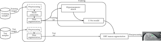
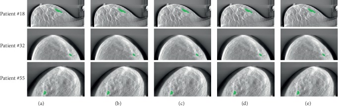
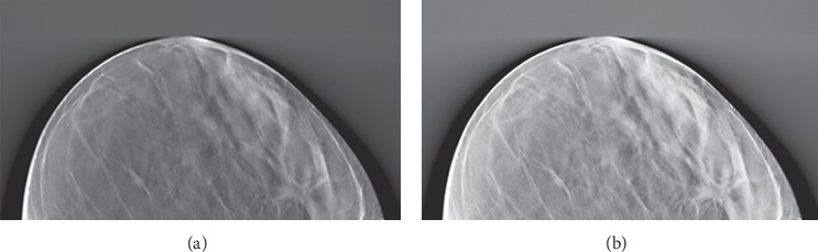
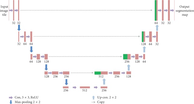
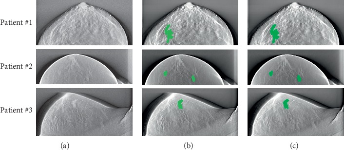
To improve the automatic segmentation accuracy of breast masses in digital breast tomosynthesis (DBT) images, we propose a DBT mass automatic segmentation algorithm by using a U-Net architecture. Firstly, to suppress the background tissue noise and enhance the contrast of the mass candidate regions, after the top-hat transform of DBT images, a constraint matrix is constructed and multiplied with the DBT image. Secondly, an efficient U-Net neural network is built and image patches are extracted before data augmentation to establish the training dataset to train the U-Net model. And then the presegmentation of the DBT tumors is implemented, which initially classifies per pixel into two different types of labels. Finally, all regions smaller than 50 voxels considered as false positives are removed, and the median filter smoothes the mass boundaries to obtain the final segmentation results. The proposed method can effectively improve the performance in the automatic segmentation of the masses in DBT images. Using the detection Accuracy (Acc), Sensitivity (Sen), Specificity (Spe), and area under the curve (AUC) as evaluation indexes, the Acc, Sen, Spe, and AUC for DBT mass segmentation in the entire experimental dataset is 0.871, 0.869, 0.882, and 0.859, respectively. Our proposed U-Net-based DBT mass automatic segmentation system obtains promising results, which is superior to some classical architectures, and may be expected to have clinical application prospects.

摘要

为了提高数字乳腺断层合成(DBT)图像中乳腺肿块的自动分割精度,我们提出了一种基于 U-Net 架构的 DBT 肿块自动分割算法。首先,通过对 DBT 图像进行顶帽变换,抑制背景组织噪声,增强肿块候选区域的对比度,构建约束矩阵并与 DBT 图像相乘。其次,构建一个高效的 U-Net 神经网络,并在数据增强之前提取图像补丁,以建立训练数据集来训练 U-Net 模型。然后对 DBT 肿瘤进行预分割,初步将每个像素分为两种不同类型的标签。最后,去除所有小于 50 体素的区域作为假阳性,并通过中值滤波器平滑肿块边界,得到最终的分割结果。该方法可以有效地提高 DBT 图像中肿块自动分割的性能。使用检测准确率(Acc)、灵敏度(Sen)、特异性(Spe)和曲线下面积(AUC)作为评价指标,整个实验数据集的 DBT 肿块分割的 Acc、Sen、Spe 和 AUC 分别为 0.871、0.869、0.882 和 0.859。我们提出的基于 U-Net 的 DBT 肿块自动分割系统取得了有前景的结果,优于一些经典的架构,有望具有临床应用前景。

https://cdn.ncbi.nlm.nih.gov/pmc/blobs/2adb/7204342/2ff79e575cca/CMMM2020-7156165.005.jpg
https://cdn.ncbi.nlm.nih.gov/pmc/blobs/2adb/7204342/39b69d74929c/CMMM2020-7156165.001.jpg
https://cdn.ncbi.nlm.nih.gov/pmc/blobs/2adb/7204342/7ffbcfd0cbc7/CMMM2020-7156165.002.jpg
https://cdn.ncbi.nlm.nih.gov/pmc/blobs/2adb/7204342/e0682f5bcd3b/CMMM2020-7156165.003.jpg
https://cdn.ncbi.nlm.nih.gov/pmc/blobs/2adb/7204342/b2bf5dfc0a90/CMMM2020-7156165.004.jpg
https://cdn.ncbi.nlm.nih.gov/pmc/blobs/2adb/7204342/2ff79e575cca/CMMM2020-7156165.005.jpg

相似文献

1
DBT Masses Automatic Segmentation Using U-Net Neural Networks.基于 U-Net 神经网络的弥散张量成像体素自动分割。
Comput Math Methods Med. 2020 Jan 28;2020:7156165. doi: 10.1155/2020/7156165. eCollection 2020.
2
Automated Segmentation of Mass Regions in DBT Images Using a Dilated DCNN Approach.基于扩张 DCNN 的乳腺断层摄影图像肿块区域自动分割。
Comput Intell Neurosci. 2022 Feb 2;2022:9082694. doi: 10.1155/2022/9082694. eCollection 2022.
3
Transfer Learning From Convolutional Neural Networks for Computer-Aided Diagnosis: A Comparison of Digital Breast Tomosynthesis and Full-Field Digital Mammography.卷积神经网络的迁移学习在计算机辅助诊断中的应用:数字乳腺断层合成与全数字化乳腺摄影的比较。
Acad Radiol. 2019 Jun;26(6):735-743. doi: 10.1016/j.acra.2018.06.019. Epub 2018 Aug 1.
4
Evaluation of a variable dose acquisition technique for microcalcification and mass detection in digital breast tomosynthesis.数字乳腺断层合成中微钙化和肿块检测的可变剂量采集技术评估
Med Phys. 2009 Jun;36(6):1976-84. doi: 10.1118/1.3116902.
5
Digital breast tomosynthesis versus digital mammography: integration of image modalities enhances deep learning-based breast mass classification.数字乳腺断层合成与数字乳腺钼靶摄影:图像模式的整合增强了基于深度学习的乳腺肿块分类。
Eur Radiol. 2020 Feb;30(2):778-788. doi: 10.1007/s00330-019-06457-5. Epub 2019 Nov 5.
6
Computerized mass detection for digital breast tomosynthesis directly from the projection images.直接从投影图像进行数字化乳腺断层合成的计算机化肿块检测。
Med Phys. 2006 Feb;33(2):482-91. doi: 10.1118/1.2163390.
7
Impact of prior mammograms on combined reading of digital mammography and digital breast tomosynthesis.既往乳腺钼靶检查对数字化乳腺钼靶摄影和数字化乳腺断层合成联合阅片的影响。
Acta Radiol. 2017 Feb;58(2):148-155. doi: 10.1177/0284185116647211. Epub 2016 Jul 20.
8
Computer-aided detection of clustered microcalcifications in digital breast tomosynthesis: a 3D approach.计算机辅助检测数字乳腺断层合成中的簇状微钙化:一种 3D 方法。
Med Phys. 2012 Jan;39(1):28-39. doi: 10.1118/1.3662072.
9
Mass detection in digital breast tomosynthesis: Deep convolutional neural network with transfer learning from mammography.数字乳腺断层合成中的肿块检测:基于乳腺X线摄影迁移学习的深度卷积神经网络
Med Phys. 2016 Dec;43(12):6654. doi: 10.1118/1.4967345.
10
Comparison of a stationary digital breast tomosynthesis system to magnified 2D mammography using breast tissue specimens.使用乳腺组织标本对固定式数字乳腺断层合成系统与放大二维乳腺摄影进行比较。
Acad Radiol. 2014 Dec;21(12):1547-52. doi: 10.1016/j.acra.2014.07.009. Epub 2014 Aug 27.

引用本文的文献

1
Deep Learning in Digital Breast Tomosynthesis: Current Status, Challenges, and Future Trends.数字乳腺断层合成中的深度学习:现状、挑战与未来趋势。
MedComm (2020). 2025 Jun 9;6(6):e70247. doi: 10.1002/mco2.70247. eCollection 2025 Jun.
2
Deep learning, radiomics and radiogenomics applications in the digital breast tomosynthesis: a systematic review.深度学习、放射组学和放射基因组学在数字乳腺断层合成中的应用:系统评价。
BMC Bioinformatics. 2023 Oct 26;24(1):401. doi: 10.1186/s12859-023-05515-6.
3
An Efficient Deep Neural Network to Classify Large 3D Images With Small Objects.

本文引用的文献

1
Fully convolutional architecture vs sliding-window CNN for corneal endothelium cell segmentation.用于角膜内皮细胞分割的全卷积架构与滑动窗口卷积神经网络对比
BMC Biomed Eng. 2019 Jan 30;1:4. doi: 10.1186/s42490-019-0003-2. eCollection 2019.
2
Breast Cancer Diagnosis in Digital Breast Tomosynthesis: Effects of Training Sample Size on Multi-Stage Transfer Learning Using Deep Neural Nets.数字乳腺断层合成中的乳腺癌诊断:使用深度神经网络的多阶段迁移学习对训练样本大小的影响。
IEEE Trans Med Imaging. 2019 Mar;38(3):686-696. doi: 10.1109/TMI.2018.2870343.
3
Optical Coherence Tomography Vulnerable Plaque Segmentation Based on Deep Residual U-Net.
一种高效的深度学习神经网络,用于对具有小物体的大型 3D 图像进行分类。
IEEE Trans Med Imaging. 2024 Jan;43(1):351-365. doi: 10.1109/TMI.2023.3302799. Epub 2024 Jan 2.
4
A Competition, Benchmark, Code, and Data for Using Artificial Intelligence to Detect Lesions in Digital Breast Tomosynthesis.人工智能用于检测数字乳腺断层合成图像中病变的竞赛、基准、代码和数据。
JAMA Netw Open. 2023 Feb 1;6(2):e230524. doi: 10.1001/jamanetworkopen.2023.0524.
5
Efficient System for Delimitation of Benign and Malignant Breast Masses.用于区分乳腺良性和恶性肿块的高效系统。
Entropy (Basel). 2022 Dec 5;24(12):1775. doi: 10.3390/e24121775.
6
YOLOv4-Based CNN Model versus Nested Contours Algorithm in the Suspicious Lesion Detection on the Mammography Image: A Direct Comparison in the Real Clinical Settings.基于YOLOv4的卷积神经网络模型与嵌套轮廓算法在乳腺钼靶图像可疑病变检测中的比较:真实临床环境下的直接对比
J Imaging. 2022 Mar 24;8(4):88. doi: 10.3390/jimaging8040088.
7
Mass Detection and Segmentation in Digital Breast Tomosynthesis Using 3D-Mask Region-Based Convolutional Neural Network: A Comparative Analysis.基于3D掩码区域卷积神经网络的数字乳腺断层合成中的肿块检测与分割:对比分析
Front Mol Biosci. 2020 Nov 11;7:599333. doi: 10.3389/fmolb.2020.599333. eCollection 2020.
基于深度残差 U-Net 的光学相干断层成像易损斑块分割。
Rev Cardiovasc Med. 2019 Sep 30;20(3):171-177. doi: 10.31083/j.rcm.2019.03.5201.
4
Artificial Intelligence for Mammography and Digital Breast Tomosynthesis: Current Concepts and Future Perspectives.人工智能在乳腺 X 线摄影和数字乳腺断层合成中的应用:现状与未来展望。
Radiology. 2019 Nov;293(2):246-259. doi: 10.1148/radiol.2019182627. Epub 2019 Sep 24.
5
Tumor Characteristics and Molecular Subtypes in Breast Cancer Screening with Digital Breast Tomosynthesis: The Malmö Breast Tomosynthesis Screening Trial.数字化乳腺断层合成筛查乳腺癌的肿瘤特征和分子亚型:马尔默乳腺断层合成筛查试验。
Radiology. 2019 Nov;293(2):273-281. doi: 10.1148/radiol.2019190132. Epub 2019 Sep 3.
6
Evaluation of a new image reconstruction method for digital breast tomosynthesis: effects on the visibility of breast lesions and breast density.数字乳腺断层合成新技术图像重建方法的评估:对乳腺病变和乳腺密度可见度的影响。
Br J Radiol. 2019 Nov;92(1103):20190345. doi: 10.1259/bjr.20190345. Epub 2019 Sep 5.
7
Technical Note: Noise models for virtual clinical trials of digital breast tomosynthesis.技术说明:数字乳腺断层合成虚拟临床试验的噪声模型。
Med Phys. 2019 Jun;46(6):2683-2689. doi: 10.1002/mp.13534. Epub 2019 May 3.
8
Double reading of automated breast ultrasound with digital mammography or digital breast tomosynthesis for breast cancer screening.自动乳腺超声与数字乳腺钼靶或数字乳腺断层融合成像用于乳腺癌筛查的双读片。
Clin Imaging. 2019 May-Jun;55:119-125. doi: 10.1016/j.clinimag.2019.01.019. Epub 2019 Jan 23.
9
Automatic Breast and Fibroglandular Tissue Segmentation in Breast MRI Using Deep Learning by a Fully-Convolutional Residual Neural Network U-Net.基于深度学习的全卷积残差神经网络 U-Net 在乳腺 MRI 中自动分割乳腺和纤维腺体组织。
Acad Radiol. 2019 Nov;26(11):1526-1535. doi: 10.1016/j.acra.2019.01.012. Epub 2019 Jan 31.
10
Comparison of two-view digital breast tomosynthesis to three-view digital mammography in a simulated screening setting.在模拟筛查环境下,双视图数字乳腺断层合成与三视图数字乳腺摄影的比较。
Acta Radiol. 2019 Sep;60(9):1094-1101. doi: 10.1177/0284185118815315. Epub 2018 Dec 10.