IEEE Trans Med Imaging. 2024 Jan;43(1):351-365. doi: 10.1109/TMI.2023.3302799. Epub 2024 Jan 2.
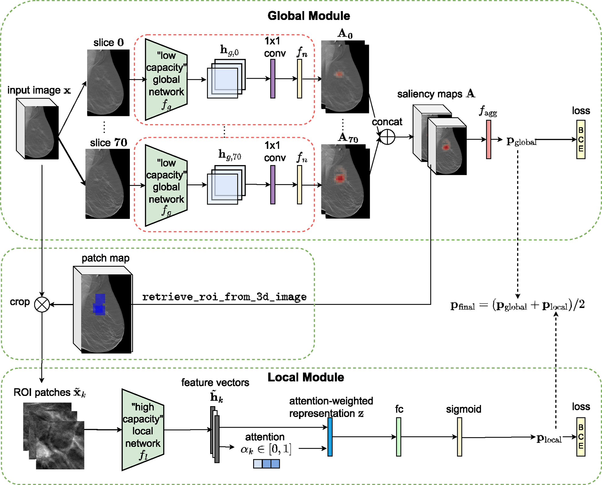
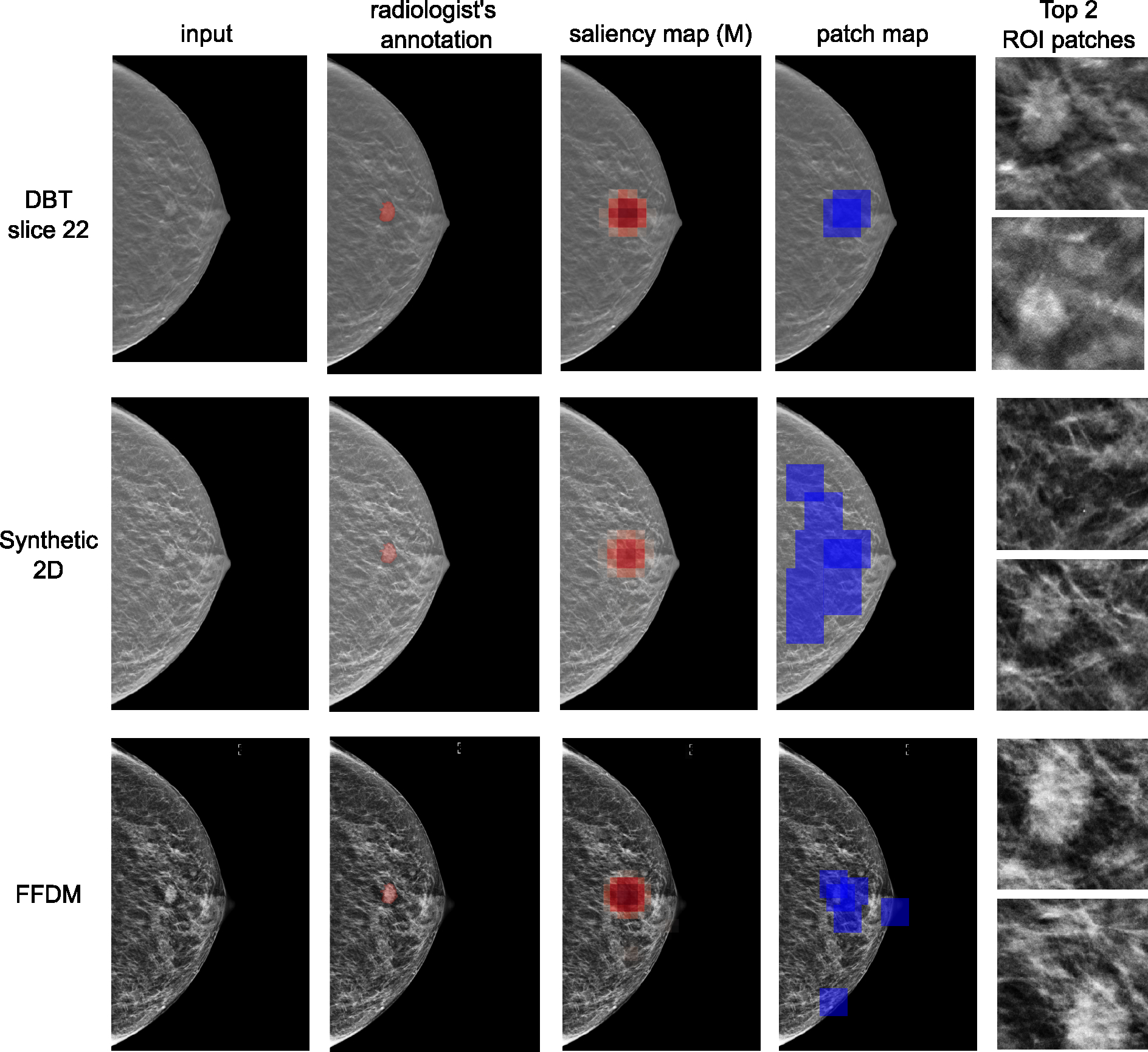
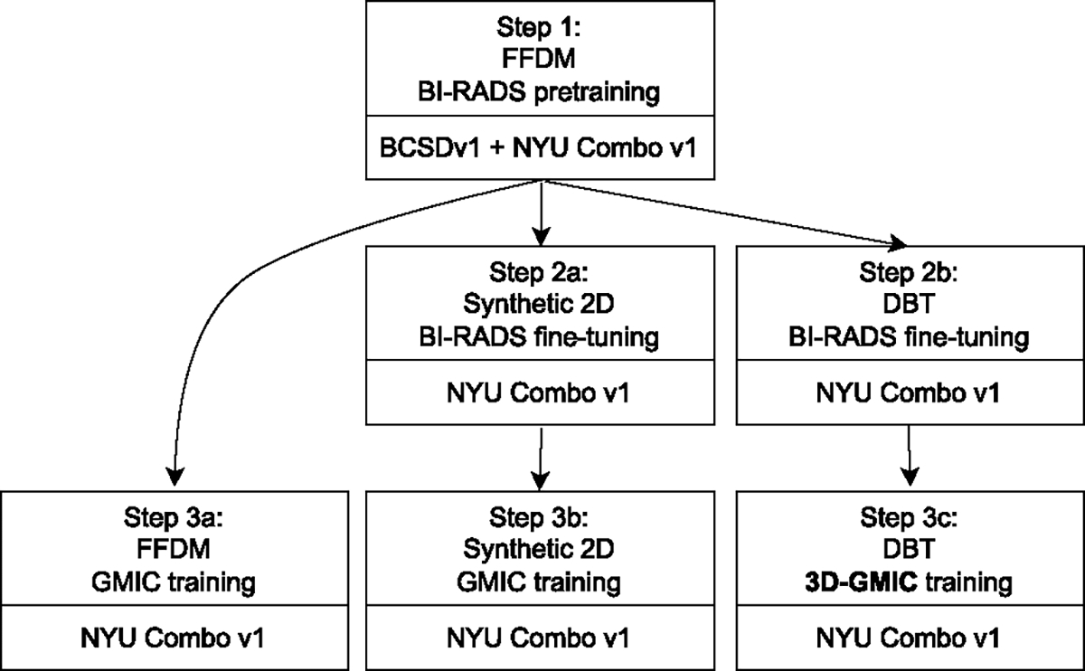
3D imaging enables accurate diagnosis by providing spatial information about organ anatomy. However, using 3D images to train AI models is computationally challenging because they consist of 10x or 100x more pixels than their 2D counterparts. To be trained with high-resolution 3D images, convolutional neural networks resort to downsampling them or projecting them to 2D. We propose an effective alternative, a neural network that enables efficient classification of full-resolution 3D medical images. Compared to off-the-shelf convolutional neural networks, our network, 3D Globally-Aware Multiple Instance Classifier (3D-GMIC), uses 77.98%-90.05% less GPU memory and 91.23%-96.02% less computation. While it is trained only with image-level labels, without segmentation labels, it explains its predictions by providing pixel-level saliency maps. On a dataset collected at NYU Langone Health, including 85,526 patients with full-field 2D mammography (FFDM), synthetic 2D mammography, and 3D mammography, 3D-GMIC achieves an AUC of 0.831 (95% CI: 0.769-0.887) in classifying breasts with malignant findings using 3D mammography. This is comparable to the performance of GMIC on FFDM (0.816, 95% CI: 0.737-0.878) and synthetic 2D (0.826, 95% CI: 0.754-0.884), which demonstrates that 3D-GMIC successfully classified large 3D images despite focusing computation on a smaller percentage of its input compared to GMIC. Therefore, 3D-GMIC identifies and utilizes extremely small regions of interest from 3D images consisting of hundreds of millions of pixels, dramatically reducing associated computational challenges. 3D-GMIC generalizes well to BCS-DBT, an external dataset from Duke University Hospital, achieving an AUC of 0.848 (95% CI: 0.798-0.896).
3D 成像通过提供有关器官解剖结构的空间信息来实现准确诊断。然而,使用 3D 图像来训练人工智能模型在计算上具有挑战性,因为它们的像素比其 2D 对应物多 10 倍或 100 倍。为了使用高分辨率 3D 图像进行训练,卷积神经网络需要对其进行下采样或投影到 2D。我们提出了一种有效的替代方案,即一种能够有效分类全分辨率 3D 医学图像的神经网络。与现成的卷积神经网络相比,我们的网络 3D 全局感知多实例分类器(3D-GMIC)使用的 GPU 内存少 77.98%-90.05%,计算量少 91.23%-96.02%。虽然它仅使用图像级标签进行训练,而没有分割标签,但它通过提供像素级显着性图来解释其预测。在纽约大学朗格尼健康中心收集的一个数据集上,包括 85526 名接受全视野 2D 乳房 X 光摄影(FFDM)、合成 2D 乳房 X 光摄影和 3D 乳房 X 光摄影的患者,3D-GMIC 在使用 3D 乳房 X 光摄影对具有恶性发现的乳房进行分类时,AUC 为 0.831(95%CI:0.769-0.887)。这与 GMIC 在 FFDM(0.816,95%CI:0.737-0.878)和合成 2D(0.826,95%CI:0.754-0.884)上的性能相当,这表明 3D-GMIC 成功地对大型 3D 图像进行了分类,尽管与 GMIC 相比,它将计算重点集中在输入的一小部分上。因此,3D-GMIC 可以识别和利用由数亿像素组成的 3D 图像中的极小感兴趣区域,从而大大降低了相关的计算挑战。3D-GMIC 很好地推广到了 BCS-DBT,这是杜克大学医院的一个外部数据集,其 AUC 为 0.848(95%CI:0.798-0.896)。