Dpto Medicina Clínica, Universidad Miguel Hernández, 03550 San Juan de Alicante, Spain.
CIBERehd, Instituto de Salud Carlos III, 28029 Madrid, Spain.
Cells. 2020 May 15;9(5):1227. doi: 10.3390/cells9051227.
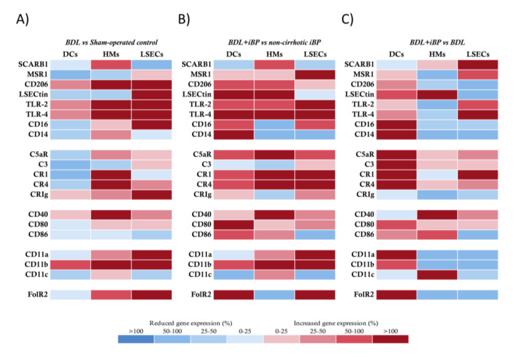
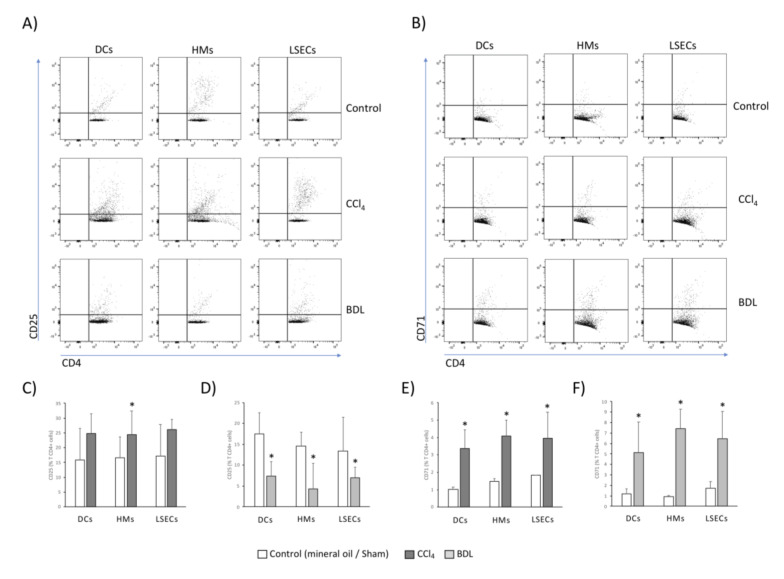
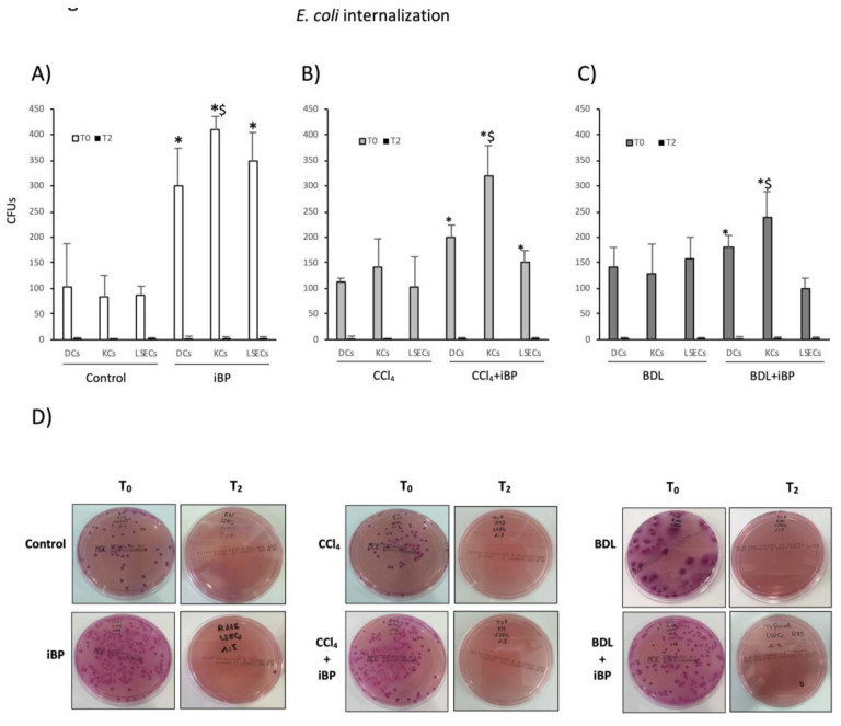
Hepatic immune function is compromised during cirrhosis. This study investigated the immune features of liver sinusoidal endothelial cells (LSECs) in two experimental models of cirrhosis. Dendritic cells, hepatic macrophages, and LSECs were isolated from carbon tetrachloride and bile duct-ligated rats. Gene expression of innate receptors, bacterial internalization, co-stimulatory molecules induction, and CD4+ T cell activation and differentiation were evaluated. Induced bacterial peritonitis and norfloxacin protocols on cirrhotic rats were also carried out. LSECs demonstrated an active immunosurveillance profile, as shown by transcriptional modulation of different scavenger and cell-adhesion genes, and their contribution to bacterial internalization. LSECs significantly increased their expression of CD40 and CD80 and stimulated CD4+ T cell activation marker CD71 in both models. The pro-inflammatory Th17 subset was expanded in CCl-derived LSECs co-cultures. In the bile duct ligation (BDL) model, CD4+ T cell differentiation only occurred under induced bacterial peritonitis conditions. Differentiated pro-inflammatory Th cells by LSECs in both experimental models were significantly reduced with norfloxacin treatment, whereas Foxp3 tolerogenic Th CD4+ cells were expanded. Conclusion: LSECs' participation in the innate-adaptive immune progression, their ability to stimulate pro-inflammatory CD4+ T cells expansion during liver damage, and their target role in norfloxacin-induced immunomodulation granted a specific competence to this cell population in cirrhosis.
肝脏免疫功能在肝硬化期间受损。本研究在两种肝硬化实验模型中研究了肝窦内皮细胞 (LSEC) 的免疫特征。从四氯化碳和胆管结扎大鼠中分离出树突状细胞、肝巨噬细胞和 LSEC。评估了先天受体的基因表达、细菌内化、共刺激分子诱导以及 CD4+T 细胞激活和分化。还对肝硬化大鼠进行了诱导性细菌性腹膜炎和诺氟沙星方案。LSEC 表现出活跃的免疫监视特征,表现为不同的清道夫和细胞黏附基因的转录调节,以及它们对细菌内化的贡献。在两种模型中,LSEC 显著增加了 CD40 和 CD80 的表达,并刺激了 CD4+T 细胞激活标志物 CD71。在 CCl 衍生的 LSEC 共培养物中,促炎性 Th17 亚群扩增。在胆管结扎 (BDL) 模型中,仅在诱导性细菌性腹膜炎条件下才发生 CD4+T 细胞分化。用诺氟沙星治疗后,两种实验模型中由 LSEC 分化的促炎性 Th 细胞显著减少,而 Foxp3 耐受诱导的 Th CD4+细胞则扩增。结论:LSEC 参与固有-适应性免疫进展,它们在肝损伤期间刺激促炎性 CD4+T 细胞扩增的能力,以及它们在诺氟沙星诱导的免疫调节中的靶作用赋予了这群细胞在肝硬化中的特定功能。