Fang Mengjie, Kan Yangyang, Dong Di, Yu Tao, Zhao Nannan, Jiang Wenyan, Zhong Lianzhen, Hu Chaoen, Luo Yahong, Tian Jie
School of Artificial Intelligence, University of Chinese Academy of Sciences, Beijing, China.
CAS Key Laboratory of Molecular Imaging, Institute of Automation, Chinese Academy of Sciences, Beijing, China.
Front Oncol. 2020 May 5;10:563. doi: 10.3389/fonc.2020.00563. eCollection 2020.
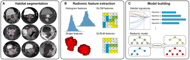
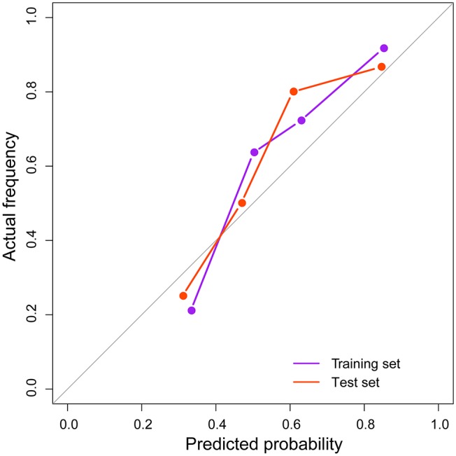
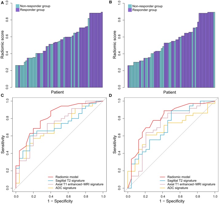
To develop a radiomic model based on multiparametric magnetic resonance imaging (MRI) for predicting treatment response prior to commencing concurrent chemotherapy and radiation therapy (CCRT) for locally advanced cervical cancer. The retrospective study enrolled 120 patients (allocated to a training or a test set) with locally advanced cervical cancer who underwent CCRT between December 2014 and June 2017. All patients enrolled underwent MRI with nine sequences before treatment and again at the end of the fourth week of treatment. Responses were evaluated by MRI according to RECIST standards, and patients were divided into a responder group or non-responder group. For every MRI sequence, a total of 114 radiomic features were extracted from the outlined tumor habitat. On the training set, the least absolute shrinkage and selection operator method was used to select key features and to construct nine habitat signatures. Then, three kinds of machine learning models were compared and applied to integrate these predictive signatures and the clinical characteristics into a radiomic model. The discrimination ability, reliability, and calibration of our radiomic model were evaluated. The radiomic model, which consisted of three habitat signatures from sagittal T2 image, axial T1 enhanced-MRI image, and ADC image, respectively, has shown good predictive performance, with area under the curve of 0.820 (95% CI: 0.713-0.927) in the training set and 0.798 (95% CI: 0.678-0.917) in the test set. Meanwhile, the model proved to perform better than each single signature or clinical characteristic. A radiomic model employing features from multiple tumor habitats held the ability for predicting treatment response in patients with locally advanced cervical cancer before commencing CCRT. These results illustrated a potential new tool for improving medical decision-making and therapeutic strategies.
基于多参数磁共振成像(MRI)开发一种放射组学模型,用于在开始局部晚期宫颈癌的同步放化疗(CCRT)之前预测治疗反应。这项回顾性研究纳入了2014年12月至2017年6月期间接受CCRT的120例局部晚期宫颈癌患者(分为训练集或测试集)。所有纳入患者在治疗前及治疗第四周结束时均接受了9个序列的MRI检查。根据RECIST标准通过MRI评估反应情况,患者被分为反应组或无反应组。对于每个MRI序列,从勾勒出的肿瘤区域中提取总共114个放射组学特征。在训练集上,使用最小绝对收缩和选择算子方法选择关键特征并构建9个区域特征。然后,比较并应用三种机器学习模型将这些预测特征和临床特征整合到一个放射组学模型中。评估了我们的放射组学模型的判别能力、可靠性和校准情况。该放射组学模型分别由矢状位T2图像、轴位T1增强MRI图像和ADC图像的三个区域特征组成,在训练集和测试集中均显示出良好的预测性能,训练集曲线下面积为0.820(95%CI:0.713 - 0.927),测试集为0.798(95%CI:0.678 - 0.917)。同时,该模型被证明比每个单一特征或临床特征表现更好。一个采用来自多个肿瘤区域特征的放射组学模型具有在开始CCRT之前预测局部晚期宫颈癌患者治疗反应的能力。这些结果说明了一种用于改善医疗决策和治疗策略的潜在新工具。