Department of Liver Surgery, Peking Union Medical College (PUMC) Hospital, PUMC & Chinese Academy of Medical Sciences, Beijing, China.
Biomanufacturing Center, Department of Mechanical Engineering, Tsinghua University, Beijing, China.
Gut. 2021 Mar;70(3):567-574. doi: 10.1136/gutjnl-2019-319960. Epub 2020 May 20.
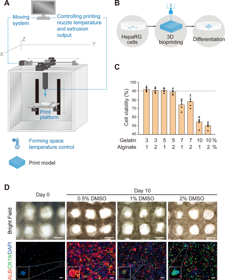
Shortage of organ donors, a critical challenge for treatment of end-stage organ failure, has motivated the development of alternative strategies to generate organs in vitro. Here, we aim to describe the hepatorganoids, which is a liver tissue model generated by three-dimensional (3D) bioprinting of HepaRG cells and investigate its liver functions in vitro and in vivo.
3D bioprinted hepatorganoids (3DP-HOs) were constructed using HepaRG cells and bioink, according to specific 3D printing procedures. Liver functions of 3DP-HOs were detected after 7 days of differentiation in vitro, which were later transplanted into Fah-deficient mice. The in vivo liver functions of 3DP-HOs were evaluated by survival time and liver damage of mice, human liver function markers and human-specific debrisoquine metabolite production.
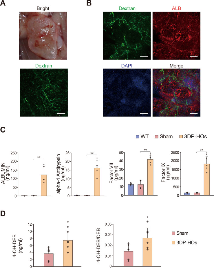
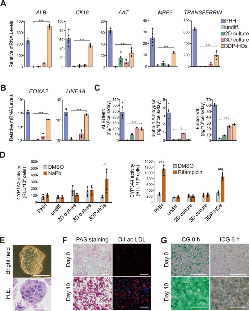
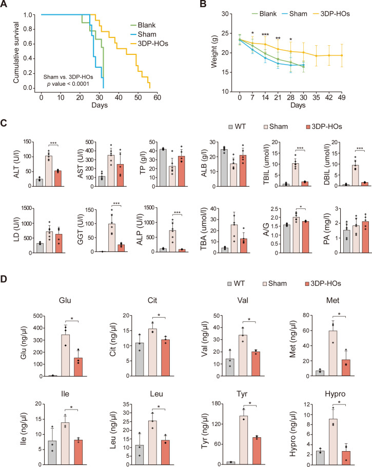
3DP-HOs broadly acquired liver functions, such as ALBUMIN secretion, drug metabolism and glycogen storage after 7 days of differentiation. After transplantation into abdominal cavity of mouse model of liver injury, 3DP-HOs further matured and displayed increased synthesis of liver-specific proteins. Particularly, the mice acquired human-specific drug metabolism activities. Functional vascular systems were also formed in transplanted 3DP-HOs, further enhancing the material transport and liver functions of 3DP-HOs. Most importantly, transplantation of 3DP-HOs significantly improved the survival of mice.
Our results demonstrated a comprehensive proof of principle, which indicated that 3DP-HO model of liver tissues possessed in vivo hepatic functions and alleviated liver failure after transplantation, suggesting that 3D bioprinting could be used to generate human liver tissues as the alternative transplantation donors for treatment of liver diseases.
器官捐献短缺是治疗终末期器官衰竭的一个关键挑战,这促使人们开发了替代策略,以在体外生成器官。本文旨在描述肝类器官,它是通过 HepaRG 细胞的三维(3D)生物打印生成的肝脏组织模型,并研究其在体外和体内的肝脏功能。
根据特定的 3D 打印程序,使用 HepaRG 细胞和生物墨水构建 3D 生物打印的肝类器官(3DP-HO)。在体外分化 7 天后检测 3DP-HO 的肝脏功能,随后将其移植到 Fah 缺陷型小鼠体内。通过小鼠的存活时间和肝损伤、人肝功能标志物和人特异性地昔洛韦代谢产物的产生来评估 3DP-HO 的体内肝脏功能。
3DP-HO 在分化 7 天后广泛获得了肝脏功能,如白蛋白分泌、药物代谢和糖原储存。在移植到肝损伤小鼠的腹腔后,3DP-HO 进一步成熟,并显示出肝脏特异性蛋白合成的增加。特别是,小鼠获得了人特异性的药物代谢活性。移植的 3DP-HO 中还形成了功能性血管系统,进一步增强了 3DP-HO 的物质转运和肝脏功能。最重要的是,移植 3DP-HO 显著提高了小鼠的存活率。
我们的结果提供了一个全面的原理验证,表明 3DP-HO 肝脏组织模型具有体内肝脏功能,并在移植后缓解了肝衰竭,这表明 3D 生物打印可用于生成人类肝脏组织,作为治疗肝脏疾病的替代移植供体。