Department of Creative IT Engineering, Pohang University of Science and Technology, Pohang, Republic of Korea.
Biomedical Engineering Research Center, Smart Healthcare Research Institute, Samsung Medical Center, Sungkyunkwan University School of Medicine, Seoul, Republic of Korea.
PLoS One. 2020 May 26;15(5):e0233531. doi: 10.1371/journal.pone.0233531. eCollection 2020.
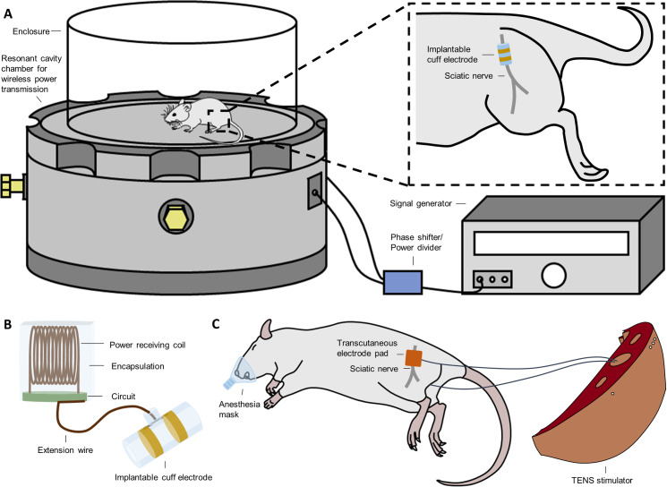
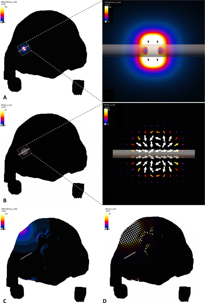
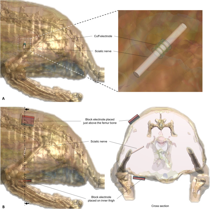
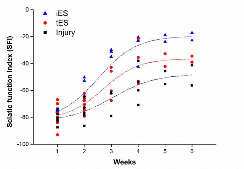
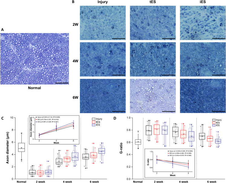
Several studies have investigated the use of invasive and non-invasive stimulation methods to enhance nerve regeneration, and varying degrees of effectiveness have been reported. However, due to the use of different parameters in these studies, a fair comparison between the effectiveness of invasive and non-invasive stimulation methods is not possible. The present study compared the effectiveness of invasive and non-invasive stimulation using similar parameters. Eighteen Sprague Dawley rats were classified into three groups: the iES group stimulated with fully implantable device, the tES group stimulated with transcutaneous electrical nerve stimulation (TENS), and the injury group (no stimulation). The iES and tES groups received stimulation for 6 weeks starting immediately after the injury. Motor function was evaluated using the sciatic functional index (SFI) every week. The SFI values increased over time in all groups; faster and superior functional recovery was observed in the iES group than in the tES group. Histological evaluation of the nerve sections and gastrocnemius muscle sections were performed every other week. The axon diameter and muscle fiber area in the iES group were larger, and the g-ratio in the iES group was closer to 0.6 than those in the tES group. To assess the cause of the difference in efficiency, a 3D rat anatomical model was used to simulate the induced electric fields in each group. A significantly higher concentration and intensity around the sciatic nerve was observed in the iES group than in the tES group. Vector field distribution showed that the field was orthogonal to the sciatic nerve spread in the tES group, whereas it was parallel in the iES group; this suggested that the tES group was less effective in nerve stimulation. The results indicated that even though rats in the TENS group showed better recovery than those in the injury group, it cannot replace direct stimulation yet because rats stimulated with the invasive method showed faster recovery and superior outcomes. This was likely attributable to the greater concentration and parallel distribution of electric field with respect to target nerve.
已有多项研究调查了使用侵入性和非侵入性刺激方法来增强神经再生的效果,报告显示其效果各有不同。然而,由于这些研究中使用了不同的参数,因此无法对侵入性和非侵入性刺激方法的效果进行公平比较。本研究使用类似的参数比较了侵入性和非侵入性刺激的效果。将 18 只 Sprague Dawley 大鼠分为三组:iES 组(使用完全可植入装置进行刺激)、tES 组(经皮电神经刺激(TENS)刺激)和损伤组(无刺激)。iES 和 tES 组在损伤后立即开始接受为期 6 周的刺激。每周使用坐骨神经功能指数(SFI)评估运动功能。所有组的 SFI 值随时间增加;iES 组的功能恢复更快、更优。每隔一周对神经切片和比目鱼肌切片进行组织学评估。iES 组的轴突直径和肌纤维面积较大,iES 组的 g-ratio 更接近 0.6,优于 tES 组。为了评估效率差异的原因,使用 3D 大鼠解剖模型模拟每组的感应电场。iES 组在坐骨神经周围观察到更高的浓度和强度,而 tES 组则没有。矢量场分布表明,tES 组中的场在坐骨神经传播中呈正交分布,而 iES 组中的场则呈平行分布;这表明 tES 组在神经刺激方面效果较差。结果表明,尽管 TENS 组的大鼠比损伤组的大鼠恢复得更好,但它不能替代直接刺激,因为使用侵入性方法刺激的大鼠恢复更快,效果更好。这可能归因于目标神经的电场浓度更高且分布更平行。