The Medical Center of Stomatology, The First Affiliated Hospital of Jinan University, Guangzhou 510630, China.
Guanghua School of Stomatology, Guangdong Provincial Key Laboratory of Stomatology, Stomatological Hospital, Sun Yat-sen University, Guangzhou 510055, China.
Biomed Res Int. 2020 May 9;2020:4671989. doi: 10.1155/2020/4671989. eCollection 2020.
This study is aimed at evaluating the effects of platelet-rich plasma (PRP) on proliferation, viability, and odontogenic differentiation of neural crest stem-like cells (NCSCs) derived from human dental apical papilla.
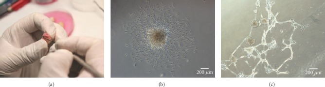
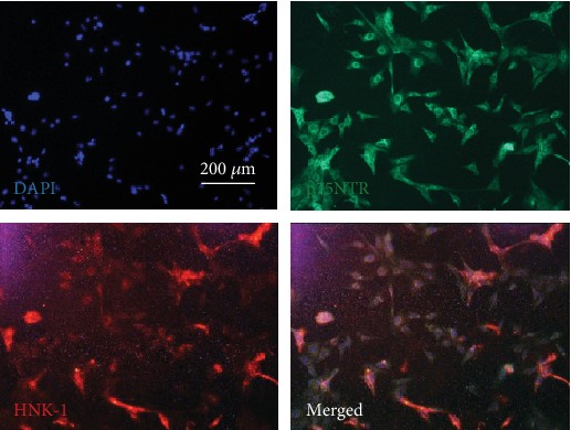
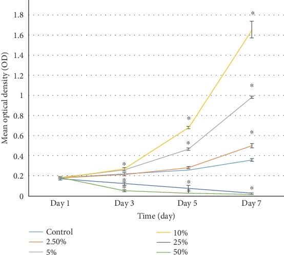
Cells from apical papillae were obtained and then induced to form neural spheres. The expression of NCSC markers p75NTR and HNK-1 in neural sphere cells was detected by immunofluorescence staining. Human PRP was prepared by a 2-step centrifugation method and activated by CaCl and thrombin. The concentrations of PDGF-BB and TGF-1 in whole blood and PRP were measured by an ELISA kit. PRP in five different concentrations (0%, 2.5%, 5%, 10%, and 25%) was applied to culture NCSCs. On the 1, 3, 5, and 7 days, cell proliferation was evaluated by CCK8. Cell viability was tested by a live/dead staining kit. mRNA and protein expression of DSPP and BMP4 were analyzed by RT-qPCR and western blot, respectively. Statistical analysis was performed by a one-way analysis of variance (ANOVA) test or -test.
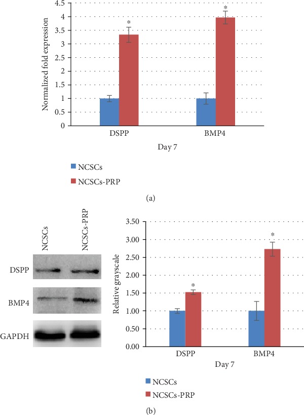
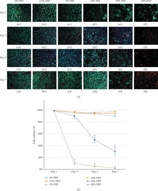
Dental apical papilla cells formed neural spheres, from which cells displayed positive expression of p75NTR and HNK-1. The concentrations of PDGF-BB and TGF-1 in PRP were about 3.5-fold higher than those in whole blood. 5% and 10% PRP significantly promoted proliferation of NCSCs, while 25% and 50% PRP inhibited cell proliferation from Day 3 to Day 7. Low-concentration (2.5%, 5%, and 10%) PRP slightly improved viability of NCSCs on Day 7. On the other hand, high-concentration (25% and 50%) PRP significantly inhibited viability of NCSCs from Day 3 to Day 7. RT-qPCR and western blot results indicated that 10% PRP could promote odontogenic differentiation of NCSCs on Day 7. mRNA and protein expression of DSPP and BMP4 were significantly upregulated in the 10% PRP group compared to those in the control group ( < 0.05).
PRP is a simply acquirable blood derivative which contains high concentration of growth factors like PDGF-BB and TGF-1. PRP in a proper concentration could promote proliferation, viability, and odontogenic differentiation of NCSCs derived from human dental apical papilla.
本研究旨在评估富血小板血浆(PRP)对人牙髓根尖乳头来源神经嵴干细胞样细胞(NCSCs)增殖、活力和牙向分化的影响。
从根尖乳头中获得细胞,然后诱导形成神经球。通过免疫荧光染色检测神经球细胞中 NCSC 标志物 p75NTR 和 HNK-1 的表达。采用两步离心法制备人 PRP,并通过 CaCl 和凝血酶激活。通过 ELISA 试剂盒测量全血和 PRP 中 PDGF-BB 和 TGF-1 的浓度。将 PRP 应用于培养 NCSCs,浓度分别为 0%、2.5%、5%、10%和 25%。在第 1、3、5 和 7 天,通过 CCK8 评估细胞增殖。通过活/死染色试剂盒检测细胞活力。通过 RT-qPCR 和 Western blot 分别分析 DSPP 和 BMP4 的 mRNA 和蛋白表达。通过单因素方差(ANOVA)检验或 t 检验进行统计分析。
牙髓根尖乳头细胞形成神经球,其中细胞显示 p75NTR 和 HNK-1 的阳性表达。PRP 中 PDGF-BB 和 TGF-1 的浓度约为全血的 3.5 倍。5%和 10% PRP 显著促进 NCSCs 的增殖,而 25%和 50% PRP 从第 3 天到第 7 天抑制细胞增殖。低浓度(2.5%、5%和 10%)PRP 可略微提高第 7 天 NCSCs 的活力。另一方面,高浓度(25%和 50%)PRP 从第 3 天到第 7 天显著抑制 NCSCs 的活力。RT-qPCR 和 Western blot 结果表明,第 7 天 10% PRP 可促进 NCSCs 的牙向分化。与对照组相比,10% PRP 组的 DSPP 和 BMP4 的 mRNA 和蛋白表达均显著上调(<0.05)。
PRP 是一种简单获得的血液衍生物,其中含有高浓度的生长因子,如 PDGF-BB 和 TGF-1。适当浓度的 PRP 可促进人牙髓根尖乳头来源 NCSCs 的增殖、活力和牙向分化。