• 文献检索
  • 文档翻译
  • 深度研究
  • 学术资讯
  • Suppr Zotero 插件Zotero 插件
  • 邀请有礼
  • 套餐&价格
  • 历史记录
应用&插件
Suppr Zotero 插件Zotero 插件浏览器插件Mac 客户端Windows 客户端微信小程序
定价
高级版会员购买积分包购买API积分包
服务
文献检索文档翻译深度研究API 文档MCP 服务
关于我们
关于 Suppr公司介绍联系我们用户协议隐私条款
关注我们

Suppr 超能文献

核心技术专利:CN118964589B侵权必究
粤ICP备2023148730 号-1Suppr @ 2026

文献检索

告别复杂PubMed语法,用中文像聊天一样搜索,搜遍4000万医学文献。AI智能推荐,让科研检索更轻松。

立即免费搜索

文件翻译

保留排版,准确专业,支持PDF/Word/PPT等文件格式,支持 12+语言互译。

免费翻译文档

深度研究

AI帮你快速写综述,25分钟生成高质量综述,智能提取关键信息,辅助科研写作。

立即免费体验

与 COVID-19 相关的独特影像生物标志物有哪些?

Any unique image biomarkers associated with COVID-19?

机构信息

Department of Radiology, University of Pittsburgh, Pittsburgh, PA, 15213, USA.

Department of Biostatistics, University of Pittsburgh, Pittsburgh, PA, 15213, USA.

出版信息

Eur Radiol. 2020 Nov;30(11):6221-6227. doi: 10.1007/s00330-020-06956-w. Epub 2020 Jul 20.

DOI:10.1007/s00330-020-06956-w
PMID:32462445
原文链接:https://pmc.ncbi.nlm.nih.gov/articles/PMC7253230/
Abstract

OBJECTIVE

To define the uniqueness of chest CT infiltrative features associated with COVID-19 image characteristics as potential diagnostic biomarkers.

METHODS

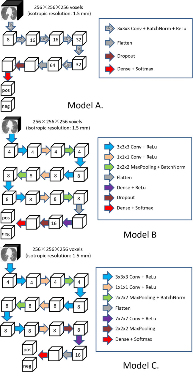
We retrospectively collected chest CT exams including n = 498 on 151 unique patients RT-PCR positive for COVID-19 and n = 497 unique patients with community-acquired pneumonia (CAP). Both COVID-19 and CAP image sets were partitioned into three groups for training, validation, and testing respectively. In an attempt to discriminate COVID-19 from CAP, we developed several classifiers based on three-dimensional (3D) convolutional neural networks (CNNs). We also asked two experienced radiologists to visually interpret the testing set and discriminate COVID-19 from CAP. The classification performance of the computer algorithms and the radiologists was assessed using the receiver operating characteristic (ROC) analysis, and the nonparametric approaches with multiplicity adjustments when necessary.

RESULTS

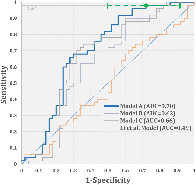
One of the considered models showed non-trivial, but moderate diagnostic ability overall (AUC of 0.70 with 99% CI 0.56-0.85). This model allowed for the identification of 8-50% of CAP patients with only 2% of COVID-19 patients.

CONCLUSIONS

Professional or automated interpretation of CT exams has a moderately low ability to distinguish between COVID-19 and CAP cases. However, the automated image analysis is promising for targeted decision-making due to being able to accurately identify a sizable subsect of non-COVID-19 cases.

KEY POINTS

• Both human experts and artificial intelligent models were used to classify the CT scans. • ROC analysis and the nonparametric approaches were used to analyze the performance of the radiologists and computer algorithms. • Unique image features or patterns may not exist for reliably distinguishing all COVID-19 from CAP; however, there may be imaging markers that can identify a sizable subset of non-COVID-19 cases.

摘要

目的

定义与 COVID-19 图像特征相关的胸部 CT 浸润特征的独特性,将其作为潜在的诊断生物标志物。

方法

我们回顾性地收集了胸部 CT 检查,包括 n=151 例经 RT-PCR 检测为 COVID-19 阳性的患者和 n=497 例社区获得性肺炎(CAP)患者的 498 次胸部 CT 检查。将 COVID-19 和 CAP 图像集分别分为三组进行训练、验证和测试。为了尝试将 COVID-19 与 CAP 区分开来,我们基于三维(3D)卷积神经网络(CNN)开发了几种分类器。我们还请两位有经验的放射科医生对测试集进行视觉解释,并将 COVID-19 与 CAP 区分开来。使用受试者工作特征(ROC)分析评估计算机算法和放射科医生的分类性能,并在必要时使用非参数方法进行多重调整。

结果

所考虑的模型之一显示出整体上具有一定但中等的诊断能力(AUC 为 0.70,99%CI 为 0.56-0.85)。该模型允许仅将 2%的 COVID-19 患者识别为 CAP 患者的 8-50%。

结论

专业或自动解读 CT 检查对区分 COVID-19 和 CAP 病例的能力较低。然而,由于能够准确识别相当大一部分非 COVID-19 病例,自动图像分析具有很大的潜力用于有针对性的决策。

关键点

  • 专家和人工智能模型都用于对 CT 扫描进行分类。

  • 使用 ROC 分析和非参数方法来分析放射科医生和计算机算法的性能。

  • 可能不存在可靠地区分所有 COVID-19 与 CAP 的独特图像特征或模式;但是,可能存在能够识别相当大一部分非 COVID-19 病例的成像标志物。

https://cdn.ncbi.nlm.nih.gov/pmc/blobs/655a/7253230/c472f7d7d187/330_2020_6956_Fig2_HTML.jpg
https://cdn.ncbi.nlm.nih.gov/pmc/blobs/655a/7253230/fac394c2f963/330_2020_6956_Fig1_HTML.jpg
https://cdn.ncbi.nlm.nih.gov/pmc/blobs/655a/7253230/c472f7d7d187/330_2020_6956_Fig2_HTML.jpg
https://cdn.ncbi.nlm.nih.gov/pmc/blobs/655a/7253230/fac394c2f963/330_2020_6956_Fig1_HTML.jpg
https://cdn.ncbi.nlm.nih.gov/pmc/blobs/655a/7253230/c472f7d7d187/330_2020_6956_Fig2_HTML.jpg

相似文献

1
Any unique image biomarkers associated with COVID-19?与 COVID-19 相关的独特影像生物标志物有哪些?
Eur Radiol. 2020 Nov;30(11):6221-6227. doi: 10.1007/s00330-020-06956-w. Epub 2020 Jul 20.
2
Using Artificial Intelligence to Detect COVID-19 and Community-acquired Pneumonia Based on Pulmonary CT: Evaluation of the Diagnostic Accuracy.基于肺部 CT 的人工智能检测 COVID-19 和社区获得性肺炎:诊断准确性评估。
Radiology. 2020 Aug;296(2):E65-E71. doi: 10.1148/radiol.2020200905. Epub 2020 Mar 19.
3
Artificial Intelligence Augmentation of Radiologist Performance in Distinguishing COVID-19 from Pneumonia of Other Origin at Chest CT.人工智能增强放射科医生在胸部 CT 上区分 COVID-19 与其他病因肺炎的性能。
Radiology. 2020 Sep;296(3):E156-E165. doi: 10.1148/radiol.2020201491. Epub 2020 Apr 27.
4
Dual-Sampling Attention Network for Diagnosis of COVID-19 From Community Acquired Pneumonia.双采样注意网络用于诊断社区获得性肺炎中的 COVID-19。
IEEE Trans Med Imaging. 2020 Aug;39(8):2595-2605. doi: 10.1109/TMI.2020.2995508.
5
From community-acquired pneumonia to COVID-19: a deep learning-based method for quantitative analysis of COVID-19 on thick-section CT scans.从社区获得性肺炎到 COVID-19:一种基于深度学习的 CT 厚层扫描 COVID-19 定量分析方法。
Eur Radiol. 2020 Dec;30(12):6828-6837. doi: 10.1007/s00330-020-07042-x. Epub 2020 Jul 18.
6
Deep Learning COVID-19 Features on CXR Using Limited Training Data Sets.基于有限训练数据集的 X 光胸片深度学习 COVID-19 特征
IEEE Trans Med Imaging. 2020 Aug;39(8):2688-2700. doi: 10.1109/TMI.2020.2993291. Epub 2020 May 8.
7
A model based on CT radiomic features for predicting RT-PCR becoming negative in coronavirus disease 2019 (COVID-19) patients.基于 CT 影像组学特征的模型预测 2019 冠状病毒病(COVID-19)患者 RT-PCR 转为阴性。
BMC Med Imaging. 2020 Oct 20;20(1):118. doi: 10.1186/s12880-020-00521-z.
8
End-to-end automatic differentiation of the coronavirus disease 2019 (COVID-19) from viral pneumonia based on chest CT.基于胸部 CT 的 2019 年冠状病毒病(COVID-19)与病毒性肺炎的端到端自动区分。
Eur J Nucl Med Mol Imaging. 2020 Oct;47(11):2516-2524. doi: 10.1007/s00259-020-04929-1. Epub 2020 Jun 22.
9
COVID-19 on Chest Radiographs: A Multireader Evaluation of an Artificial Intelligence System.COVID-19 胸片:人工智能系统的多读者评估。
Radiology. 2020 Sep;296(3):E166-E172. doi: 10.1148/radiol.2020201874. Epub 2020 May 8.
10
A Weakly-Supervised Framework for COVID-19 Classification and Lesion Localization From Chest CT.一种基于弱监督的 COVID-19 分类和胸部 CT 病变定位框架。
IEEE Trans Med Imaging. 2020 Aug;39(8):2615-2625. doi: 10.1109/TMI.2020.2995965.

引用本文的文献

1
AFEX-Net: Adaptive feature extraction convolutional neural network for classifying computerized tomography images.AFEX-Net:用于对计算机断层扫描图像进行分类的自适应特征提取卷积神经网络。
Digit Health. 2024 Feb 22;10:20552076241232882. doi: 10.1177/20552076241232882. eCollection 2024 Jan-Dec.
2
Computed tomography-based radiomics improves non-invasive diagnosis of Pneumocystis jirovecii pneumonia in non-HIV patients: a retrospective study.基于计算机断层扫描的放射组学可提高非 HIV 患者肺孢子菌肺炎的无创诊断:一项回顾性研究。
BMC Pulm Med. 2024 Jan 2;24(1):11. doi: 10.1186/s12890-023-02827-4.
3
Automatic diagnosis of COVID-19 from CT images using CycleGAN and transfer learning.

本文引用的文献

1
The indispensable role of chest CT in the detection of coronavirus disease 2019 (COVID-19).胸部CT在2019冠状病毒病(COVID-19)检测中的不可或缺作用。
Eur J Nucl Med Mol Imaging. 2020 Jul;47(7):1638-1639. doi: 10.1007/s00259-020-04795-x. Epub 2020 Apr 3.
2
Chest CT Features of COVID-19 in Rome, Italy.意大利罗马地区 COVID-19 的胸部 CT 特征。
Radiology. 2020 Aug;296(2):E79-E85. doi: 10.1148/radiol.2020201237. Epub 2020 Apr 3.
3
A role for CT in COVID-19? What data really tell us so far.CT在新冠病毒肺炎中的作用?目前数据究竟告诉了我们什么。
使用循环生成对抗网络(CycleGAN)和迁移学习从CT图像中自动诊断新型冠状病毒肺炎(COVID-19)
Appl Soft Comput. 2023 Sep;144:110511. doi: 10.1016/j.asoc.2023.110511. Epub 2023 Jun 13.
4
Harnessing Machine Learning in Early COVID-19 Detection and Prognosis: A Comprehensive Systematic Review.利用机器学习进行新冠肺炎早期检测与预后:一项全面的系统综述。
Cureus. 2023 May 1;15(5):e38373. doi: 10.7759/cureus.38373. eCollection 2023 May.
5
A Review of the Machine Learning Algorithms for Covid-19 Case Analysis.用于新冠病例分析的机器学习算法综述
IEEE Trans Artif Intell. 2022 Jan 11;4(1):44-59. doi: 10.1109/TAI.2022.3142241. eCollection 2023 Feb.
6
Biomarkers: Promising and valuable tools towards diagnosis, prognosis and treatment of Covid-19 and other diseases.生物标志物:用于新冠病毒病及其他疾病诊断、预后评估和治疗的有前景且有价值的工具。
Heliyon. 2023 Feb;9(2):e13323. doi: 10.1016/j.heliyon.2023.e13323. Epub 2023 Jan 30.
7
Towards smart diagnostic methods for COVID-19: Review of deep learning for medical imaging.迈向COVID-19的智能诊断方法:医学成像深度学习综述
IPEM Transl. 2022 Nov-Dec;3:100008. doi: 10.1016/j.ipemt.2022.100008. Epub 2022 Oct 26.
8
Artificial intelligence and IoT based prediction of Covid-19 using chest X-ray images.基于人工智能和物联网,利用胸部X光图像预测新冠病毒肺炎
Smart Health (Amst). 2022 Sep;25:100299. doi: 10.1016/j.smhl.2022.100299. Epub 2022 Jun 26.
9
Current Artificial Intelligence (AI) Techniques, Challenges, and Approaches in Controlling and Fighting COVID-19: A Review.当前人工智能 (AI) 技术在控制和抗击 COVID-19 方面的挑战和方法:综述。
Int J Environ Res Public Health. 2022 May 12;19(10):5901. doi: 10.3390/ijerph19105901.
10
Machine Learning-Based Research for COVID-19 Detection, Diagnosis, and Prediction: A Survey.基于机器学习的COVID-19检测、诊断和预测研究:一项综述。
SN Comput Sci. 2022;3(4):286. doi: 10.1007/s42979-022-01184-z. Epub 2022 May 12.
Lancet. 2020 Apr 11;395(10231):1189-1190. doi: 10.1016/S0140-6736(20)30728-5. Epub 2020 Mar 27.
4
The role of CT in case ascertainment and management of COVID-19 pneumonia in the UK: insights from high-incidence regions.CT在英国2019冠状病毒病肺炎病例确诊及管理中的作用:来自高发病地区的见解
Lancet Respir Med. 2020 May;8(5):438-440. doi: 10.1016/S2213-2600(20)30132-6. Epub 2020 Mar 25.
5
Imaging Features of Coronavirus disease 2019 (COVID-19): Evaluation on Thin-Section CT.2019 年冠状病毒病(COVID-19)的影像学特征:薄层 CT 评估。
Acad Radiol. 2020 May;27(5):609-613. doi: 10.1016/j.acra.2020.03.002. Epub 2020 Mar 20.
6
Performance of Radiologists in Differentiating COVID-19 from Non-COVID-19 Viral Pneumonia at Chest CT.放射科医生在胸部 CT 鉴别 COVID-19 与非 COVID-19 病毒性肺炎中的表现。
Radiology. 2020 Aug;296(2):E46-E54. doi: 10.1148/radiol.2020200823. Epub 2020 Mar 10.
7
Clinical and High-Resolution CT Features of the COVID-19 Infection: Comparison of the Initial and Follow-up Changes.COVID-19 感染的临床和高分辨率 CT 特征:初始与随访变化比较。
Invest Radiol. 2020 Jun;55(6):332-339. doi: 10.1097/RLI.0000000000000674.
8
CT Features of Coronavirus Disease 2019 (COVID-19) Pneumonia in 62 Patients in Wuhan, China.中国武汉 62 例 2019 年冠状病毒病(COVID-19)肺炎的 CT 特征。
AJR Am J Roentgenol. 2020 Jun;214(6):1287-1294. doi: 10.2214/AJR.20.22975. Epub 2020 Mar 5.
9
COVID-19 pneumonia: what has CT taught us?新型冠状病毒肺炎:CT 给了我们哪些启示?
Lancet Infect Dis. 2020 Apr;20(4):384-385. doi: 10.1016/S1473-3099(20)30134-1. Epub 2020 Feb 24.
10
Radiological findings from 81 patients with COVID-19 pneumonia in Wuhan, China: a descriptive study.中国武汉 81 例新冠肺炎患者的放射学特征:一项描述性研究。
Lancet Infect Dis. 2020 Apr;20(4):425-434. doi: 10.1016/S1473-3099(20)30086-4. Epub 2020 Feb 24.