• 文献检索
  • 文档翻译
  • 深度研究
  • 学术资讯
  • Suppr Zotero 插件Zotero 插件
  • 邀请有礼
  • 套餐&价格
  • 历史记录
应用&插件
Suppr Zotero 插件Zotero 插件浏览器插件Mac 客户端Windows 客户端微信小程序
定价
高级版会员购买积分包购买API积分包
服务
文献检索文档翻译深度研究API 文档MCP 服务
关于我们
关于 Suppr公司介绍联系我们用户协议隐私条款
关注我们

Suppr 超能文献

核心技术专利:CN118964589B侵权必究
粤ICP备2023148730 号-1Suppr @ 2026

文献检索

告别复杂PubMed语法,用中文像聊天一样搜索,搜遍4000万医学文献。AI智能推荐,让科研检索更轻松。

立即免费搜索

文件翻译

保留排版,准确专业,支持PDF/Word/PPT等文件格式,支持 12+语言互译。

免费翻译文档

深度研究

AI帮你快速写综述,25分钟生成高质量综述,智能提取关键信息,辅助科研写作。

立即免费体验

高分辨率光学切片成像用于可视化人类耳甲艇的真皮动脉和神经

High-Resolution Episcopic Imaging for Visualization of Dermal Arteries and Nerves of the Auricular Cymba Conchae in Humans.

作者信息

Dabiri Babak, Kampusch Stefan, Geyer Stefan H, Le Van Hoang, Weninger Wolfgang J, Széles Jozsef Constantin, Kaniusas Eugenijus

机构信息

Institute of Electrodynamics, Microwave and Circuit Engineering, Vienna University of Technology, Vienna, Austria.

SzeleSTIM GmbH, Vienna, Austria.

出版信息

Front Neuroanat. 2020 May 12;14:22. doi: 10.3389/fnana.2020.00022. eCollection 2020.

DOI:10.3389/fnana.2020.00022
PMID:32477074
原文链接:https://pmc.ncbi.nlm.nih.gov/articles/PMC7236887/
Abstract

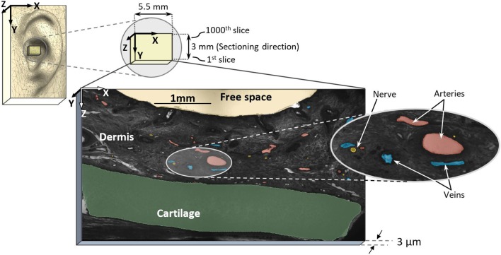
Therapeutic applications of auricular vagus nerve stimulation (VNS) have drawn recent attention. Since the targeted stimulation process and parameters depend on the electrode-tissue interaction, the lack of structural anatomical information on innervation and vascularization of the auricle restrain the current optimization of stimulation paradigms. For the first time, we employed high-resolution episcopic imaging (HREM) to generate histologic volume data from donated human cadaver ears. Optimal parameters for specimen preparation were evaluated. Anatomical 3D vascular and nerve structures were reconstructed in one sample of an auricular cymba conchae (CC). The feasibility of HREM to visualize anatomical structures was assessed in that diameters, occupied areas, volumes, and mutual distances between auricular arteries, nerves, and veins were registered. The selected region of CC (3 × 5.5 mm) showed in its cross-sections 21.7 ± 2.7 (mean ± standard deviation) arteries and 14.66 ± 2.74 nerve fibers. Identified nerve diameters were 33.66 ± 21.71 μm, and arteries had diameters in the range of 71.58 ± 80.70 μm. The respective occupied area showed a share of, on average, 2.71% and 0.3% for arteries and nerves, respectively, and similar volume occupancy for arteries and nerves. Inter-centroid minimum distance between arteries and nerves was 274 ± 222 μm. The density of vessels and nerves around a point within CC on a given grid was assessed, showing that 50% of all vessels and nerves were found in a radial distance of 1.6-1.8 mm from any of these points, which is strategically relevant when using stimulation needles in the auricle for excitation of nerves. HREM seems suitable for anatomical studies of the human ear. A 3D model of CC was established in the micrometer scale, which forms the basis for future optimization of the auricular VNS. Obviously, the presented single cadaver study needs to be validated by additional anatomical data on the innervation and vascularization of the auricle.

摘要

耳迷走神经刺激(VNS)的治疗应用近来受到关注。由于靶向刺激过程和参数取决于电极与组织的相互作用,缺乏关于耳廓神经支配和血管形成的结构解剖信息限制了当前刺激模式的优化。我们首次采用高分辨率表面成像(HREM)从捐赠的人类尸体耳朵生成组织学体积数据。评估了标本制备的最佳参数。在一个耳甲艇(CC)样本中重建了解剖学三维血管和神经结构。通过记录耳廓动脉、神经和静脉之间的直径、占据面积、体积和相互距离,评估了HREM可视化解剖结构的可行性。所选的CC区域(3×5.5毫米)在其横截面上显示有21.7±2.7(平均值±标准差)条动脉和14.66±2.74条神经纤维。识别出的神经直径为33.66±21.71微米,动脉直径在71.58±80.70微米范围内。动脉和神经各自的占据面积平均分别为2.71%和0.3%,动脉和神经的体积占有率相似。动脉和神经之间的质心最小距离为274±222微米。评估了给定网格上CC内一点周围血管和神经的密度,结果表明,所有血管和神经的50%位于距这些点中任何一点径向距离1.6 - 1.8毫米处,这在使用耳廓刺激针激发神经时具有重要的战略意义。HREM似乎适用于人类耳朵的解剖学研究。在微米尺度上建立了CC的三维模型,这为未来耳廓VNS的优化奠定了基础。显然,所呈现的单一尸体研究需要通过关于耳廓神经支配和血管形成的额外解剖数据进行验证。

https://cdn.ncbi.nlm.nih.gov/pmc/blobs/cf44/7236887/530c0c965f6d/fnana-14-00022-g0009.jpg
https://cdn.ncbi.nlm.nih.gov/pmc/blobs/cf44/7236887/15cc49617b23/fnana-14-00022-g0001.jpg
https://cdn.ncbi.nlm.nih.gov/pmc/blobs/cf44/7236887/6c1b1dab32c7/fnana-14-00022-g0002.jpg
https://cdn.ncbi.nlm.nih.gov/pmc/blobs/cf44/7236887/17833f395751/fnana-14-00022-g0003.jpg
https://cdn.ncbi.nlm.nih.gov/pmc/blobs/cf44/7236887/9ea9337ee50c/fnana-14-00022-g0004.jpg
https://cdn.ncbi.nlm.nih.gov/pmc/blobs/cf44/7236887/14a80e38a9bd/fnana-14-00022-g0005.jpg
https://cdn.ncbi.nlm.nih.gov/pmc/blobs/cf44/7236887/ebb711a16a71/fnana-14-00022-g0006.jpg
https://cdn.ncbi.nlm.nih.gov/pmc/blobs/cf44/7236887/b2838f4479c6/fnana-14-00022-g0007.jpg
https://cdn.ncbi.nlm.nih.gov/pmc/blobs/cf44/7236887/1a77da555982/fnana-14-00022-g0008.jpg
https://cdn.ncbi.nlm.nih.gov/pmc/blobs/cf44/7236887/530c0c965f6d/fnana-14-00022-g0009.jpg

相似文献

1
High-Resolution Episcopic Imaging for Visualization of Dermal Arteries and Nerves of the Auricular Cymba Conchae in Humans.高分辨率光学切片成像用于可视化人类耳甲艇的真皮动脉和神经
Front Neuroanat. 2020 May 12;14:22. doi: 10.3389/fnana.2020.00022. eCollection 2020.
2
Innervation of the Human Cavum Conchae and Auditory Canal: Anatomical Basis for Transcutaneous Auricular Nerve Stimulation.人类鼻甲腔和耳道的神经支配:经皮耳神经刺激的解剖学基础。
Biomed Res Int. 2017;2017:7830919. doi: 10.1155/2017/7830919. Epub 2017 Mar 15.
3
Transcutaneous Auricular Vagus Nerve Stimulation.经皮耳迷走神经刺激。
J Clin Neurophysiol. 2019 Nov;36(6):437-442. doi: 10.1097/WNP.0000000000000576.
4
Tragus or cymba conchae? Investigating the anatomical foundation of transcutaneous auricular vagus nerve stimulation (taVNS).耳屏还是耳甲艇?探究经皮耳迷走神经刺激(taVNS)的解剖学基础。
Brain Stimul. 2018 Jul-Aug;11(4):947-948. doi: 10.1016/j.brs.2018.06.003. Epub 2018 Jun 6.
5
Non-invasive Access to the Vagus Nerve Central Projections via Electrical Stimulation of the External Ear: fMRI Evidence in Humans.通过电刺激外耳对迷走神经中枢投射进行非侵入性通路研究:人类功能磁共振成像证据
Brain Stimul. 2015 May-Jun;8(3):624-36. doi: 10.1016/j.brs.2014.11.018. Epub 2014 Dec 6.
6
Anatomical and technical aspects of harvesting the auricle as a neurovascular facial subunit transplant in humans.将耳廓作为神经血管性面部亚单位移植用于人体的解剖学及技术方面。
Plast Reconstr Surg. 2007 Nov;120(6):1540-1545. doi: 10.1097/01.prs.0000287992.28125.ce.
7
[Effect of electroacupuncture of different regions of the auricle on epileptic seizures in epilepsy rats].[耳郭不同部位电针对癫痫大鼠癫痫发作的影响]
Zhen Ci Yan Jiu. 2011 Dec;36(6):414-8.
8
Numerical modeling of percutaneous auricular vagus nerve stimulation: a realistic 3D model to evaluate sensitivity of neural activation to electrode position.经皮耳迷走神经刺激的数值建模:评估神经激活对电极位置敏感性的真实 3D 模型。
Med Biol Eng Comput. 2017 Oct;55(10):1763-1772. doi: 10.1007/s11517-017-1629-7. Epub 2017 Feb 13.
9
Networks of Nerve Fibers, and Blood and Lymphatic Vessels in the Mouse Auricle: The Structural Basis of Ear Acupuncture.小鼠耳廓中的神经纤维、血管和淋巴管网络:耳针疗法的结构基础。
Med Acupunct. 2024 Apr 1;36(2):79-86. doi: 10.1089/acu.2023.0077. Epub 2024 Apr 11.
10
Transcutaneous electrical stimulation at auricular acupoints innervated by auricular branch of vagus nerve pairing tone for tinnitus: study protocol for a randomized controlled clinical trial.经皮电刺激迷走神经耳支支配的耳穴并结合音调治疗耳鸣:一项随机对照临床试验的研究方案
Trials. 2015 Mar 19;16:101. doi: 10.1186/s13063-015-0630-4.

引用本文的文献

1
Clinical Efficacy of Auricular Vagus Nerve Stimulation in the Treatment of Chronic and Acute Pain: A Systematic Review and Meta-analysis.耳迷走神经刺激治疗慢性和急性疼痛的临床疗效:一项系统评价和荟萃分析
Pain Ther. 2024 Dec;13(6):1407-1427. doi: 10.1007/s40122-024-00657-8. Epub 2024 Oct 9.
2
Imaging of the vascular distribution of the outer ear using optical coherence tomography angiography for highly accurate positioning of a hearable sensor.使用光学相干断层扫描血管造影术对外耳血管分布进行成像,以实现可听传感器的高精度定位。
APL Bioeng. 2024 May 23;8(2):026113. doi: 10.1063/5.0203582. eCollection 2024 Jun.
3
Networks of Nerve Fibers, and Blood and Lymphatic Vessels in the Mouse Auricle: The Structural Basis of Ear Acupuncture.

本文引用的文献

1
Stimulation Pattern Efficiency in Percutaneous Auricular Vagus Nerve Stimulation: Experimental Versus Numerical Data.经皮耳迷走神经刺激刺激模式效率:实验与数值数据比较。
IEEE Trans Biomed Eng. 2020 Jul;67(7):1921-1935. doi: 10.1109/TBME.2019.2950777. Epub 2019 Oct 31.
2
Current Directions in the Auricular Vagus Nerve Stimulation I - A Physiological Perspective.耳迷走神经刺激的当前研究方向I——生理学视角
Front Neurosci. 2019 Aug 9;13:854. doi: 10.3389/fnins.2019.00854. eCollection 2019.
3
Current Directions in the Auricular Vagus Nerve Stimulation II - An Engineering Perspective.
小鼠耳廓中的神经纤维、血管和淋巴管网络:耳针疗法的结构基础。
Med Acupunct. 2024 Apr 1;36(2):79-86. doi: 10.1089/acu.2023.0077. Epub 2024 Apr 11.
4
Randomized controlled study to evaluate the safety and clinical impact of percutaneous auricular vagus nerve stimulation in patients with severe COVID-19.评估经皮耳迷走神经刺激对重症新型冠状病毒肺炎患者安全性和临床影响的随机对照研究。
Front Physiol. 2023 Aug 8;14:1223347. doi: 10.3389/fphys.2023.1223347. eCollection 2023.
5
Immediate effects and duration of a short and single application of transcutaneous auricular vagus nerve stimulation on P300 event related potential.经皮耳迷走神经刺激单次短暂应用对P300事件相关电位的即时效应及持续时间
Front Neurosci. 2023 Mar 27;17:1096865. doi: 10.3389/fnins.2023.1096865. eCollection 2023.
6
Quantitative Image Processing for Three-Dimensional Episcopic Images of Biological Structures: Current State and Future Directions.生物结构三维光学切片图像的定量图像处理:现状与未来方向
Biomedicines. 2023 Mar 15;11(3):909. doi: 10.3390/biomedicines11030909.
7
Mapping the functional anatomy and topography of the cardiac autonomic innervation for selective cardiac neuromodulation using MicroCT.利用微型计算机断层扫描(MicroCT)绘制心脏自主神经支配的功能解剖结构和地形图,以进行选择性心脏神经调节。
Front Cell Dev Biol. 2022 Sep 12;10:968870. doi: 10.3389/fcell.2022.968870. eCollection 2022.
8
Percutaneous Auricular Vagus Nerve Stimulation Reduces Inflammation in Critical Covid-19 Patients.经皮耳迷走神经刺激可减轻重症新冠肺炎患者的炎症反应。
Front Physiol. 2022 Jul 4;13:897257. doi: 10.3389/fphys.2022.897257. eCollection 2022.
9
Auricular vagus nerve stimulator for closed-loop biofeedback-based operation.用于基于闭环生物反馈操作的耳迷走神经刺激器。
Analog Integr Circuits Signal Process. 2022;112(2):237-246. doi: 10.1007/s10470-022-02037-8. Epub 2022 May 10.
10
Functional anatomy of the vagus system - Emphasis on the somato-visceral interface.迷走神经系统的功能解剖学——重点关注躯体-内脏界面。
Auton Neurosci. 2021 Dec;236:102887. doi: 10.1016/j.autneu.2021.102887. Epub 2021 Sep 28.
耳迷走神经刺激的当前发展方向II——工程学视角
Front Neurosci. 2019 Jul 24;13:772. doi: 10.3389/fnins.2019.00772. eCollection 2019.
4
The Tomato and Regulate Leaf Axil Patterning Along the Proximal-Distal Axes.番茄并沿近端-远端轴调节叶腋模式。
Front Plant Sci. 2018 Aug 6;9:1126. doi: 10.3389/fpls.2018.01126. eCollection 2018.
5
Comparative study of regenerative effects of mesenchymal stem cells derived from placental amnion, chorion and umbilical cord on dermal wounds.胎盘羊膜、绒毛膜和脐带间充质干细胞对皮肤创面再生作用的比较研究。
Placenta. 2018 May;65:37-46. doi: 10.1016/j.placenta.2018.04.004. Epub 2018 Apr 10.
6
Tragus or cymba conchae? Investigating the anatomical foundation of transcutaneous auricular vagus nerve stimulation (taVNS).耳屏还是耳甲艇?探究经皮耳迷走神经刺激(taVNS)的解剖学基础。
Brain Stimul. 2018 Jul-Aug;11(4):947-948. doi: 10.1016/j.brs.2018.06.003. Epub 2018 Jun 6.
7
Transcutaneous nerve stimulation via the tragus: are we really stimulating the vagus nerve?经耳屏的经皮神经刺激:我们真的在刺激迷走神经吗?
Brain Stimul. 2018 Jul-Aug;11(4):945-946. doi: 10.1016/j.brs.2018.03.018. Epub 2018 Apr 5.
8
The dermal arteries in the cutaneous angiosome of the descending genicular artery.膝降动脉皮穿支血管的皮肤血管体中的皮肤动脉。
J Anat. 2018 Jun;232(6):979-986. doi: 10.1111/joa.12792. Epub 2018 Feb 14.
9
Auricular Neuromodulation: The Emerging Concept beyond the Stimulation of Vagus and Trigeminal Nerves.耳神经调节:超越迷走神经和三叉神经刺激的新兴概念。
Medicines (Basel). 2018 Jan 21;5(1):10. doi: 10.3390/medicines5010010.
10
High-resolution Episcopic Microscopy (HREM) - Simple and Robust Protocols for Processing and Visualizing Organic Materials.高分辨率光学显微镜(HREM)——处理和可视化有机材料的简单且可靠的方案。
J Vis Exp. 2017 Jul 7(125):56071. doi: 10.3791/56071.