Department of Urology, The People's Hospital of Guangxi Zhuang Autonomous Region, Nanning, China.
Graduate School, Guangxi Medical University, Nanning, China.
BMC Urol. 2020 Jun 1;20(1):61. doi: 10.1186/s12894-020-00635-0.
It is well known that androgen-deprivation therapy (ADT) can inevitably drive prostate cancer (PCa) cells into a castration-resistant state. According to the "Warburg effect", the metabolism of aggressive tumor cells increases significantly. The growth of cancer cells depends on glycolysis, which may be a potential target for cancer control. 6-Phosphofructo-2-kinase/fructose-2,6-biphosphatase 4 (PFKFB4) plays key roles in the proliferation and metastasis of PCa cells. However, there is very limited knowledge on the role of PFKFB4 in the conversion to castration resistance. The present study aimed to determine the changes in glucose consumption and PFKFB4 expression in LNCaP cells and androgen-independent LNCaP (LNCaP-AI) cells during the whole process of androgen-independent growth. Additionally, PFKFB4 expression in human PCa tissues was evaluated.
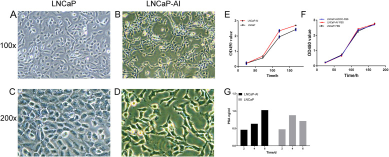
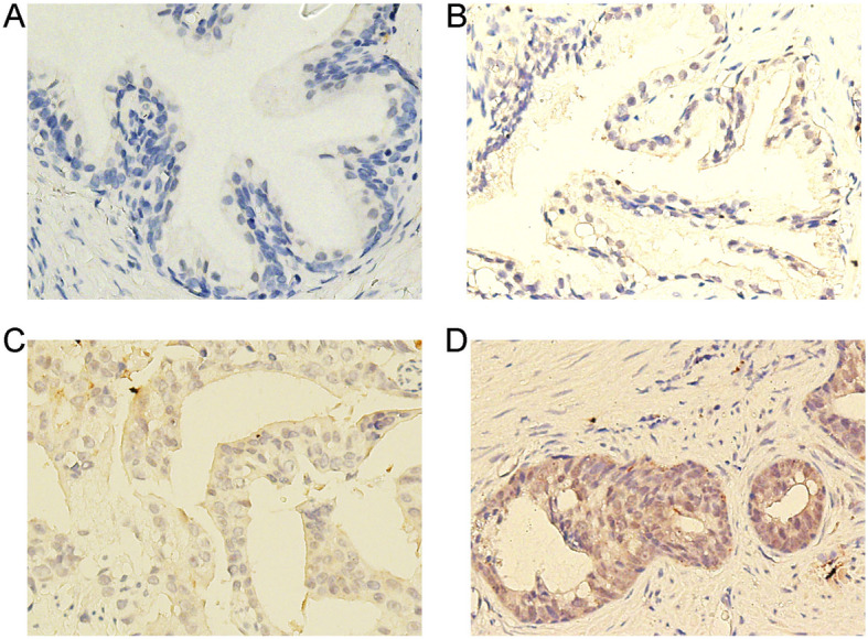
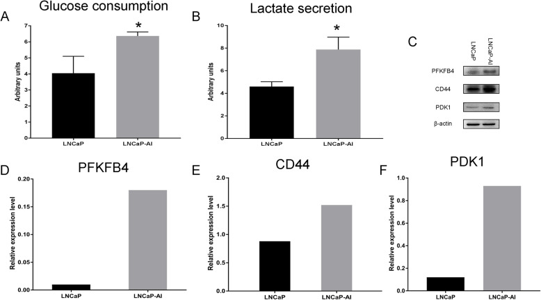
We established an androgen-independent LNCaP-AI cell line derived from LNCaP cells to mimic the traits of castration resistance in vitro. LNCaP-AI and LNCaP cells were cultured in the corresponding medium containing the same amount of glucose. At the end of experiments, the medium supernatant and blank medium were collected, and absorbance was measured. LNCaP-AI and LNCaP cells were harvested to detect PFKFB4 expression by Western blotting. Prostate tissue samples including PCa tissue, carcinoma-adjacent tissue and benign prostatic hyperplasia (BPH) tissue specimens were evaluated for PFKFB4 expression using immunohistochemistry.
In 18 h supernatant samples, the glucose consumption and lactate secretion of LNCaP-AI cells were higher than those of LNCaP cells. The Western blot results indicated that PFKFB4 expression was increased in LNCaP-AI cells compared with LNCaP cells. Immunohistochemistry revealed that the expression of PFKFB4 in PCa tissue specimens was higher than that in BPH and adjacent tissue specimens. However, the differences in PCa tissue before and after ADT were not statistically significant.
PFKFB4 may be associated with enhanced glycolysis during the androgen-independent growth of PCa cells in vitro. PFKFB4 may be a marker of PCa progression. Our results provide a rationale for further clinical investigation of PCa treatment focused on controlling PFKFB4 expression.
众所周知,去势治疗(ADT)不可避免地会使前列腺癌(PCa)细胞进入去势抵抗状态。根据“Warburg 效应”,侵袭性肿瘤细胞的代谢显著增加。癌细胞的生长依赖于糖酵解,这可能是癌症控制的一个潜在靶点。6-磷酸果糖-2-激酶/果糖-2,6-二磷酸酶 4(PFKFB4)在 PCa 细胞的增殖和转移中发挥关键作用。然而,关于 PFKFB4 在向去势抵抗转化中的作用的知识非常有限。本研究旨在确定 LNCaP 细胞和雄激素非依赖性 LNCaP(LNCaP-AI)细胞在整个雄激素非依赖性生长过程中葡萄糖消耗和 PFKFB4 表达的变化。此外,还评估了人 PCa 组织中 PFKFB4 的表达。
我们建立了一种源自 LNCaP 细胞的雄激素非依赖性 LNCaP-AI 细胞系,以模拟体外去势抵抗的特征。LNCaP-AI 和 LNCaP 细胞在含有相同葡萄糖量的相应培养基中培养。实验结束时,收集培养基上清液和空白培养基,测量吸光度。收集 LNCaP-AI 和 LNCaP 细胞,通过 Western blot 检测 PFKFB4 表达。使用免疫组织化学法评估前列腺组织标本(包括 PCa 组织、癌旁组织和良性前列腺增生(BPH)组织标本)中 PFKFB4 的表达。
在 18 h 上清液样本中,LNCaP-AI 细胞的葡萄糖消耗和乳酸分泌均高于 LNCaP 细胞。Western blot 结果表明,LNCaP-AI 细胞中 PFKFB4 的表达高于 LNCaP 细胞。免疫组织化学显示,PCa 组织标本中 PFKFB4 的表达高于 BPH 和癌旁组织标本。然而,ADT 前后 PCa 组织的差异无统计学意义。
PFKFB4 可能与体外 PCa 细胞雄激素非依赖性生长过程中糖酵解的增强有关。PFKFB4 可能是 PCa 进展的标志物。我们的研究结果为进一步研究针对控制 PFKFB4 表达的 PCa 治疗提供了依据。