Division of Image Guided Therapy, Department of Diagnostic Imaging, The Hospital for Sick Children, University of Toronto, 555 University Avenue, Toronto, ON, M5G 1X8, Canada.
Division of Rheumatology, Department of Paediatrics, The Hospital for Sick Children, University of Toronto, Toronto, Canada.
Pediatr Rheumatol Online J. 2020 Jun 17;18(1):52. doi: 10.1186/s12969-020-00435-8.
Sacroiliitis is commonly seen in enthesitis-related arthritis (ERA), a subtype of juvenile idiopathic arthritis (JIA). Sacroiliitis is characterized by the inflammation of the sacroiliac (SI) joints (+/- adjacent tissues). The treatment options include systemic therapy with or without corticosteroid SI joint injections. Image guided SI joint injections are frequently requested in pediatric patients with sacroiliitis. The purpose of this study was to evaluate the feasibility and efficacy of SI joint injections in children with sacroiliitis.
A retrospective study of patients referred to Interventional Radiology (IR) for SI joint corticosteroid injections (2000-2018). Clinical information was collected from Electronic Patient Charts and procedural details from PACS. Efficacy was determined clinically, by MRI, or both when available.
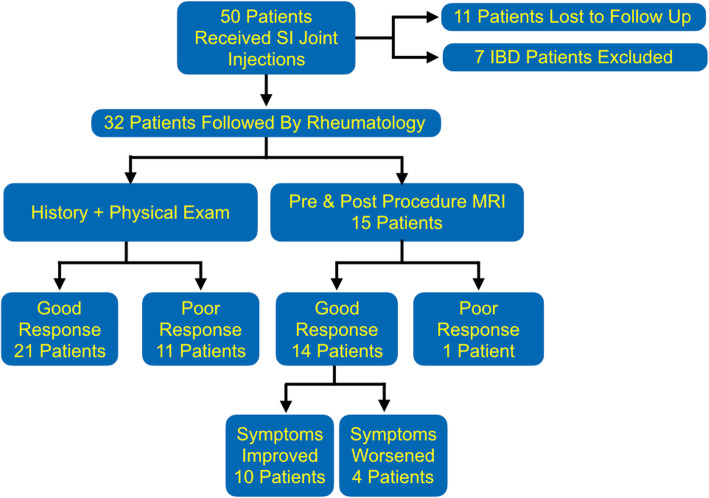
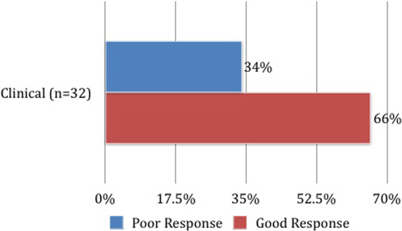
50 patients (13.8 years; M:F = 35:15) underwent image-guided SI joint corticosteroid injections. Most common indications were JIA (84%) and inflammatory bowel disease (14%). 80% had bilateral injections. 80% were performed under general anesthesia and 20% under sedation. The corticosteroid of choice was triamcinolone hexacetonide in 98% of patients. Needle guidance and confirmation was performed using CT and fluoroscopy (54%), Cone Beam CT (CBCT, 46%), with initial ultrasound assistance in 34%. All procedures were technically successful without any complications. 32/50 patients had long-term follow-up (2 years); 21/32 (66%) had clinical improvement within 3-months. Of 15 patients who had both pre- and post-procedure MRIs, 93% showed short-term improvement. At 2 years, 6% of patients were in remission, 44% continued the same treatment and 47% escalated treatment.
Image-guided SI joint injections are safe and technically feasible in children. Imaging modalities for guidance have evolved, with CBCT being the current first choice. Most patients showed short-term clinical and imaging improvement, requiring long-term maintenance or escalation of medical treatment.
骶髂关节炎常见于附着点相关关节炎(ERA),这是幼年特发性关节炎(JIA)的一个亚型。骶髂关节炎的特征是骶髂(SI)关节(+/-相邻组织)炎症。治疗选择包括全身性治疗,伴或不伴皮质类固醇骶髂关节注射。在患有骶髂关节炎的儿科患者中,经常要求进行图像引导的骶髂关节注射。本研究的目的是评估在患有骶髂关节炎的儿童中进行 SI 关节注射的可行性和疗效。
回顾性研究 2000 年至 2018 年间因 SI 关节皮质类固醇注射而转介至介入放射科的患者。临床信息从电子病历中收集,程序细节从 PACS 中收集。根据临床、MRI 或两者均有确定疗效。
50 例患者(13.8 岁;男:女=35:15)接受了图像引导的 SI 关节皮质类固醇注射。最常见的适应症是 JIA(84%)和炎症性肠病(14%)。80%为双侧注射。80%在全身麻醉下进行,20%在镇静下进行。98%的患者选择曲安奈德六乙酸酯作为皮质类固醇。96%的患者使用 CT 和透视(54%)、锥形束 CT(CBCT,46%)进行针引导和确认,最初 34%使用超声辅助。所有操作均无技术失败且无并发症。50 例患者中有 32 例(66%)获得长期随访(2 年);32 例中有 21 例(66%)在 3 个月内临床改善。15 例患者中有 15 例(93%)在接受治疗前后均行 MRI,93%显示短期改善。2 年后,6%的患者处于缓解期,44%的患者继续相同的治疗,47%的患者升级了治疗。
图像引导的 SI 关节注射在儿童中是安全且技术可行的。指导的成像方式已经发展,CBCT 是目前的首选。大多数患者表现出短期的临床和影像学改善,需要长期维持或升级药物治疗。