• 文献检索
  • 文档翻译
  • 深度研究
  • 学术资讯
  • Suppr Zotero 插件Zotero 插件
  • 邀请有礼
  • 套餐&价格
  • 历史记录
应用&插件
Suppr Zotero 插件Zotero 插件浏览器插件Mac 客户端Windows 客户端微信小程序
定价
高级版会员购买积分包购买API积分包
服务
文献检索文档翻译深度研究API 文档MCP 服务
关于我们
关于 Suppr公司介绍联系我们用户协议隐私条款
关注我们

Suppr 超能文献

核心技术专利:CN118964589B侵权必究
粤ICP备2023148730 号-1Suppr @ 2026

文献检索

告别复杂PubMed语法,用中文像聊天一样搜索,搜遍4000万医学文献。AI智能推荐,让科研检索更轻松。

立即免费搜索

文件翻译

保留排版,准确专业,支持PDF/Word/PPT等文件格式,支持 12+语言互译。

免费翻译文档

深度研究

AI帮你快速写综述,25分钟生成高质量综述,智能提取关键信息,辅助科研写作。

立即免费体验

利用人工智能(AI)鉴别海绵状血管瘤和神经鞘瘤。

Differentiate cavernous hemangioma from schwannoma with artificial intelligence (AI).

作者信息

Bi Shaowei, Chen Rongxin, Zhang Kai, Xiang Yifan, Wang Ruixin, Lin Haotian, Yang Huasheng

机构信息

State Key Laboratory of Ophthalmology, Zhongshan Ophthalmic Center, Sun Yat-sen University, Guangzhou, China.

School of Computer Science and Technology, Xidian University, Xi'an, China.

出版信息

Ann Transl Med. 2020 Jun;8(11):710. doi: 10.21037/atm.2020.03.150.

DOI:10.21037/atm.2020.03.150
PMID:32617330
原文链接:https://pmc.ncbi.nlm.nih.gov/articles/PMC7327353/
Abstract

BACKGROUND

Cavernous hemangioma and schwannoma are tumors that both occur in the orbit. Because the treatment strategies of these two tumors are different, it is necessary to distinguish them at treatment initiation. Magnetic resonance imaging (MRI) is typically used to differentiate these two tumor types; however, they present similar features in MRI images which increases the difficulty of differential diagnosis. This study aims to devise and develop an artificial intelligence framework to improve the accuracy of clinicians' diagnoses and enable more effective treatment decisions by automatically distinguishing cavernous hemangioma from schwannoma.

METHODS

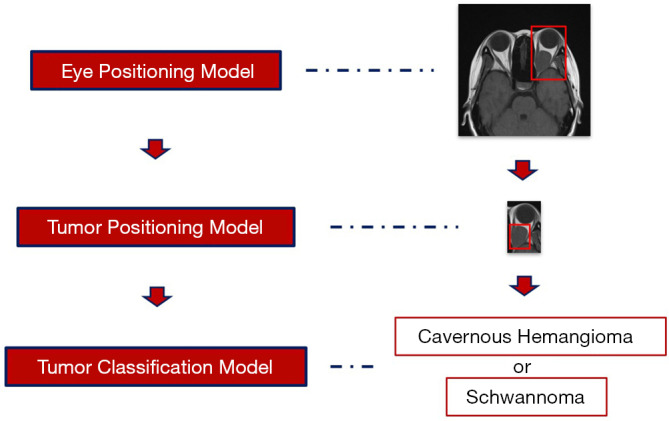
Material: As the study materials, we chose MRI images as the study materials that represented patients from diverse areas in China who had been referred to our center from more than 45 different hospitals. All images were initially acquired on films, which we scanned into digital versions and recut. Finally, 11,489 images of cavernous hemangioma (from 33 different hospitals) and 3,478 images of schwannoma (from 16 different hospitals) were collected. Labeling: All images were labeled using standard anatomical knowledge and pathological diagnosis. Training: Three types of models were trained in sequence (a total of 96 models), with each model including a specific improvement. The first two model groups were eye- and tumor-positioning models designed to reduce the identification scope, while the third model group consisted of classification models trained to make the final diagnosis.

RESULTS

First, internal four-fold cross-validation processes were conducted for all the models. During the validation of the first group, the 32 eye-positioning models were able to localize the position of the eyes with an average precision of 100%. In the second group, the 28 tumor-positioning models were able to reach an average precision above 90%. Subsequently, using the third group, the accuracy of all 32 tumor classification models reached nearly 90%. Next, external validation processes of 32 tumor classification models were conducted. The results showed that the accuracy of the transverse T1-weighted contrast-enhanced sequence reached 91.13%; the accuracy of the remaining models was significantly lower compared with the ground truth.

CONCLUSIONS

The findings of this retrospective study show that an artificial intelligence framework can achieve high accuracy, sensitivity, and specificity in automated differential diagnosis between cavernous hemangioma and schwannoma in a real-world setting, which can help doctors determine appropriate treatments.

摘要

背景

海绵状血管瘤和神经鞘瘤均为发生于眼眶的肿瘤。由于这两种肿瘤的治疗策略不同,因此在治疗开始时需要对它们进行区分。磁共振成像(MRI)通常用于区分这两种肿瘤类型;然而,它们在MRI图像中呈现出相似的特征,这增加了鉴别诊断的难度。本研究旨在设计并开发一种人工智能框架,通过自动区分海绵状血管瘤和神经鞘瘤来提高临床医生诊断的准确性,并做出更有效的治疗决策。

方法

材料:作为研究材料,我们选择MRI图像,这些图像代表了来自中国不同地区、从45多家不同医院转诊至我们中心的患者。所有图像最初是在胶片上获取的,我们将其扫描成数字版本并重新裁剪。最终,收集了11489张海绵状血管瘤图像(来自33家不同医院)和3478张神经鞘瘤图像(来自16家不同医院)。标注:所有图像均使用标准解剖学知识和病理诊断进行标注。训练:依次训练三种类型的模型(共96个模型),每个模型都有特定的改进。前两组模型是眼部和肿瘤定位模型,旨在缩小识别范围,而第三组模型是经过训练以做出最终诊断的分类模型。

结果

首先,对所有模型进行内部四折交叉验证过程。在第一组验证中,32个眼部定位模型能够定位眼睛位置,平均精度达到100%。在第二组中,28个肿瘤定位模型的平均精度达到90%以上。随后,使用第三组模型,所有32个肿瘤分类模型的准确率接近90%。接下来,对32个肿瘤分类模型进行外部验证过程。结果显示,横向T1加权对比增强序列的准确率达到91.13%;与真实情况相比,其余模型的准确率显著较低。

结论

这项回顾性研究的结果表明,人工智能框架在现实环境中对海绵状血管瘤和神经鞘瘤进行自动鉴别诊断时能够实现高准确率、敏感性和特异性,这有助于医生确定合适的治疗方案。

https://cdn.ncbi.nlm.nih.gov/pmc/blobs/1d6c/7327353/80f41e5fabe3/atm-08-11-710-f2.jpg

相似文献

1
Differentiate cavernous hemangioma from schwannoma with artificial intelligence (AI).利用人工智能(AI)鉴别海绵状血管瘤和神经鞘瘤。
Ann Transl Med. 2020 Jun;8(11):710. doi: 10.21037/atm.2020.03.150.
2
Automated Koos Classification of Vestibular Schwannoma.前庭神经鞘瘤的自动库斯分类
Front Radiol. 2022 Mar 10;2:837191. doi: 10.3389/fradi.2022.837191. eCollection 2022.
3
MRI-Based Radiomics for Differentiating Orbital Cavernous Hemangioma and Orbital Schwannoma.基于磁共振成像的影像组学用于鉴别眼眶海绵状血管瘤和眼眶神经鞘瘤
Front Med (Lausanne). 2021 Dec 16;8:795038. doi: 10.3389/fmed.2021.795038. eCollection 2021.
4
Ground truth generalizability affects performance of the artificial intelligence model in automated vertebral fracture detection on plain lateral radiographs of the spine.真实情况的可推广性会影响人工智能模型在脊柱正位侧位X线片自动检测椎体骨折中的性能。
Spine J. 2022 Apr;22(4):511-523. doi: 10.1016/j.spinee.2021.10.020. Epub 2021 Nov 1.
5
An Interpretable and Expandable Deep Learning Diagnostic System for Multiple Ocular Diseases: Qualitative Study.一种用于多种眼部疾病的可解释且可扩展的深度学习诊断系统:定性研究
J Med Internet Res. 2018 Nov 14;20(11):e11144. doi: 10.2196/11144.
6
Comparison of Chest Radiograph Interpretations by Artificial Intelligence Algorithm vs Radiology Residents.人工智能算法与放射科住院医师对胸部 X 线片解读的比较。
JAMA Netw Open. 2020 Oct 1;3(10):e2022779. doi: 10.1001/jamanetworkopen.2020.22779.
7
Differentiation of cavernous hemangioma from schwannoma of the orbit: a dynamic MRI study.眼眶海绵状血管瘤与神经鞘瘤的鉴别:动态MRI研究
AJR Am J Roentgenol. 2004 Dec;183(6):1799-804. doi: 10.2214/ajr.183.6.01831799.
8
VIIIth nerve cavernous hemangioma mimicking a stage 1 acoustic schwannoma.模仿1期听神经鞘瘤的第八脑神经海绵状血管瘤。
Neurochirurgie. 2015 Oct;61(5):352-5. doi: 10.1016/j.neuchi.2015.07.003. Epub 2015 Oct 1.
9
Differentiation between hepatic cavernous hemangioma and malignant tumor with T2-weighted MRI: comparison of fast spin-echo and breathhold fast spin-echo pulse sequences.利用T2加权磁共振成像鉴别肝海绵状血管瘤与恶性肿瘤:快速自旋回波与屏气快速自旋回波脉冲序列的比较
Clin Imaging. 1998 May-Jun;22(3):200-10. doi: 10.1016/s0899-7071(97)00124-1.
10
Artificial Intelligence System Approaching Neuroradiologist-level Differential Diagnosis Accuracy at Brain MRI.人工智能系统在脑部 MRI 中的诊断准确率接近神经放射学家水平。
Radiology. 2020 Jun;295(3):626-637. doi: 10.1148/radiol.2020190283. Epub 2020 Apr 7.

引用本文的文献

1
FaceFinder: A machine learning tool for identification of facial images from heterogenous datasets.面部识别器:一种用于从异构数据集中识别面部图像的机器学习工具。
AJO Int. 2024 Dec 11;1(4). doi: 10.1016/j.ajoint.2024.100083. Epub 2024 Nov 7.
2
The Impact of Medical Explainable Artificial Intelligence on Nurses' Innovation Behaviour: A Structural Equation Modelling Approach.医学可解释人工智能对护士创新行为的影响:一种结构方程建模方法。
J Nurs Manag. 2024 Sep 26;2024:8885760. doi: 10.1155/2024/8885760. eCollection 2024.
3
Applications of Artificial Intelligence in Ophthalmology: Glaucoma, Cornea, and Oculoplastics.

本文引用的文献

1
Prediction of cardiovascular risk factors from retinal fundus photographs via deep learning.基于深度学习的眼底图像心血管风险因素预测。
Nat Biomed Eng. 2018 Mar;2(3):158-164. doi: 10.1038/s41551-018-0195-0. Epub 2018 Feb 19.
2
Diagnosis of thyroid cancer using deep convolutional neural network models applied to sonographic images: a retrospective, multicohort, diagnostic study.使用深度卷积神经网络模型对超声图像进行甲状腺癌诊断:一项回顾性、多队列、诊断研究。
Lancet Oncol. 2019 Feb;20(2):193-201. doi: 10.1016/S1470-2045(18)30762-9. Epub 2018 Dec 21.
3
An Interpretable and Expandable Deep Learning Diagnostic System for Multiple Ocular Diseases: Qualitative Study.
人工智能在眼科中的应用:青光眼、角膜和眼整形学。
Cureus. 2024 Nov 12;16(11):e73522. doi: 10.7759/cureus.73522. eCollection 2024 Nov.
4
End-to-End Deep-Learning-Based Diagnosis of Benign and Malignant Orbital Tumors on Computed Tomography Images.基于深度学习的计算机断层扫描图像上眼眶良恶性肿瘤的端到端诊断
J Pers Med. 2023 Jan 23;13(2):204. doi: 10.3390/jpm13020204.
5
Orbital and eyelid diseases: The next breakthrough in artificial intelligence?眼眶及眼睑疾病:人工智能的下一个突破?
Front Cell Dev Biol. 2022 Nov 18;10:1069248. doi: 10.3389/fcell.2022.1069248. eCollection 2022.
6
Machine Learning for the Detection and Segmentation of Benign Tumors of the Central Nervous System: A Systematic Review.用于中枢神经系统良性肿瘤检测与分割的机器学习:一项系统综述
Cancers (Basel). 2022 May 27;14(11):2676. doi: 10.3390/cancers14112676.
一种用于多种眼部疾病的可解释且可扩展的深度学习诊断系统:定性研究
J Med Internet Res. 2018 Nov 14;20(11):e11144. doi: 10.2196/11144.
4
An Automated Grading System for Detection of Vision-Threatening Referable Diabetic Retinopathy on the Basis of Color Fundus Photographs.基于彩色眼底照片的威胁视力可转诊糖尿病视网膜病变自动分级系统。
Diabetes Care. 2018 Dec;41(12):2509-2516. doi: 10.2337/dc18-0147. Epub 2018 Oct 1.
5
Classification and mutation prediction from non-small cell lung cancer histopathology images using deep learning.基于深度学习的非小细胞肺癌组织病理学图像分类和突变预测。
Nat Med. 2018 Oct;24(10):1559-1567. doi: 10.1038/s41591-018-0177-5. Epub 2018 Sep 17.
6
Identification of Metastatic Lymph Nodes in MR Imaging with Faster Region-Based Convolutional Neural Networks.基于快速区域卷积神经网络的 MRI 中转移性淋巴结的识别。
Cancer Res. 2018 Sep 1;78(17):5135-5143. doi: 10.1158/0008-5472.CAN-18-0494. Epub 2018 Jul 19.
7
Multimodal Machine Learning: A Survey and Taxonomy.多模态机器学习:一项综述与分类法
IEEE Trans Pattern Anal Mach Intell. 2019 Feb;41(2):423-443. doi: 10.1109/TPAMI.2018.2798607. Epub 2018 Jan 25.
8
Radiological Analysis of Orbital Cavernous Hemangiomas: A Review and Comparison Between Computed Tomography and Magnetic Resonance Imaging.眼眶海绵状血管瘤的影像学分析:计算机断层扫描与磁共振成像的综述及比较
J Craniofac Surg. 2018 May;29(3):712-716. doi: 10.1097/SCS.0000000000004291.
9
Development and Validation of a Deep Learning System for Diabetic Retinopathy and Related Eye Diseases Using Retinal Images From Multiethnic Populations With Diabetes.使用来自多民族糖尿病患者群体的视网膜图像开发并验证用于糖尿病视网膜病变及相关眼病的深度学习系统
JAMA. 2017 Dec 12;318(22):2211-2223. doi: 10.1001/jama.2017.18152.
10
Optic Nerve Meningioma Mimicking Cavernous Hemangioma.酷似海绵状血管瘤的视神经脑膜瘤
World Neurosurg. 2018 Feb;110:301-302. doi: 10.1016/j.wneu.2017.11.107. Epub 2017 Nov 28.