Materials Science and Engineering Program, University of California San Diego, USA.
Department of NanoEngineering, University of California San Diego, USA.
Biomaterials. 2020 Oct;256:120204. doi: 10.1016/j.biomaterials.2020.120204. Epub 2020 Jun 22.
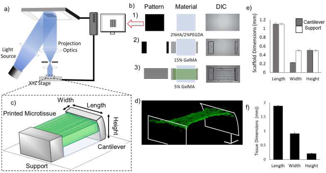
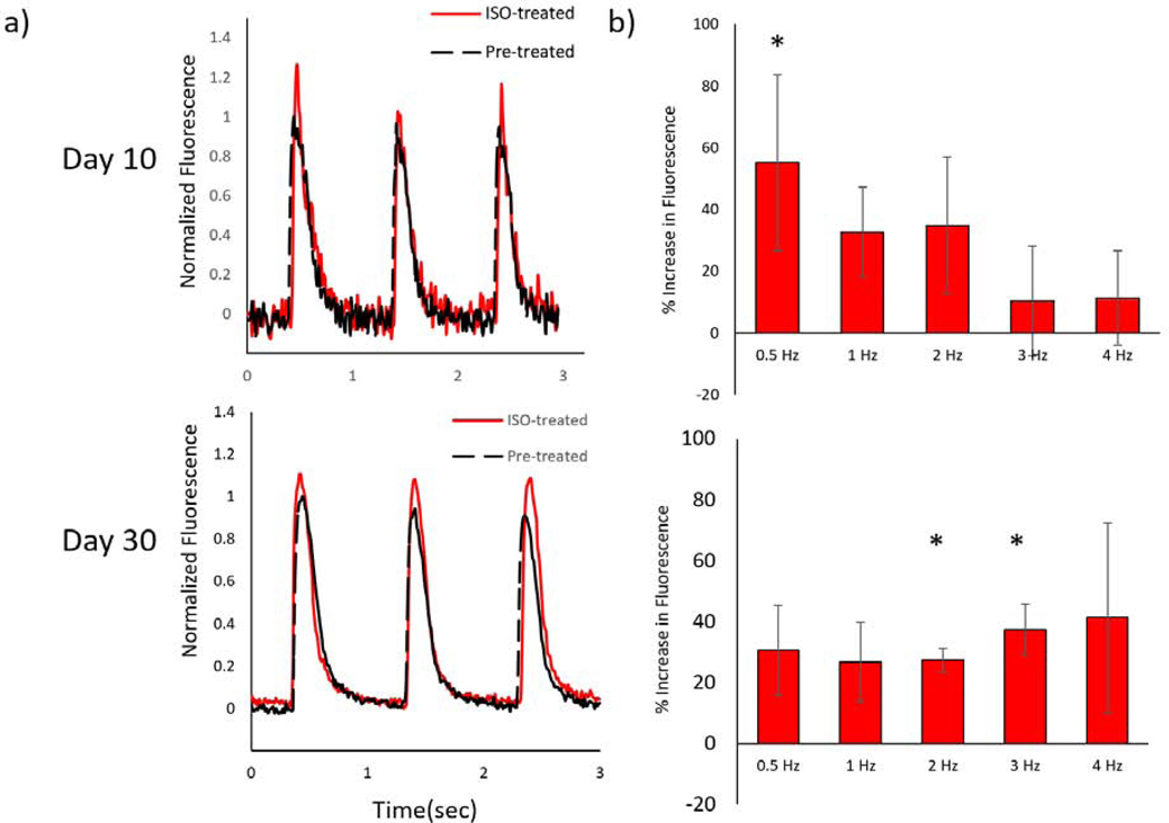
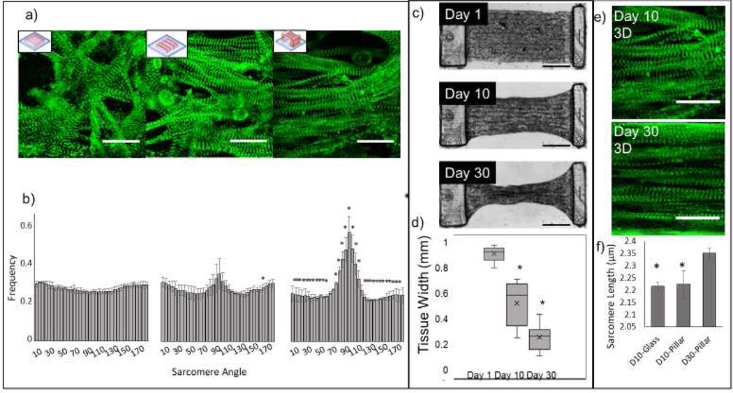
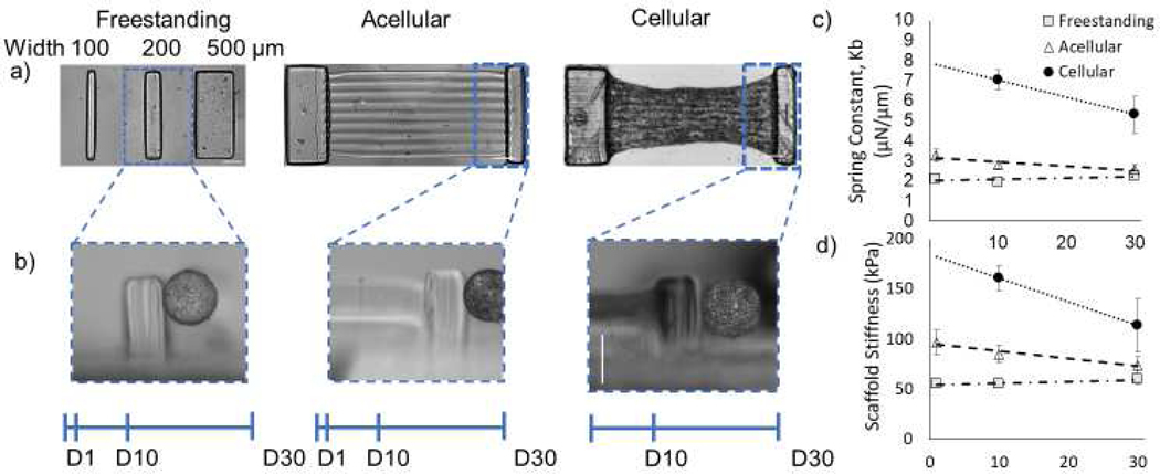
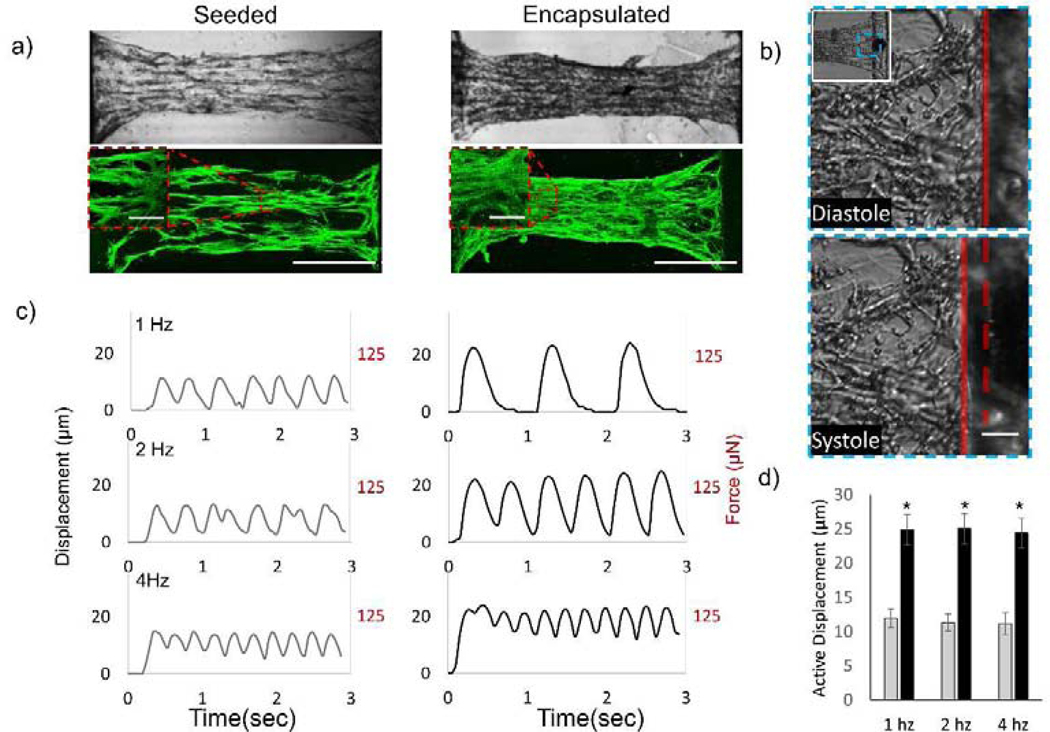
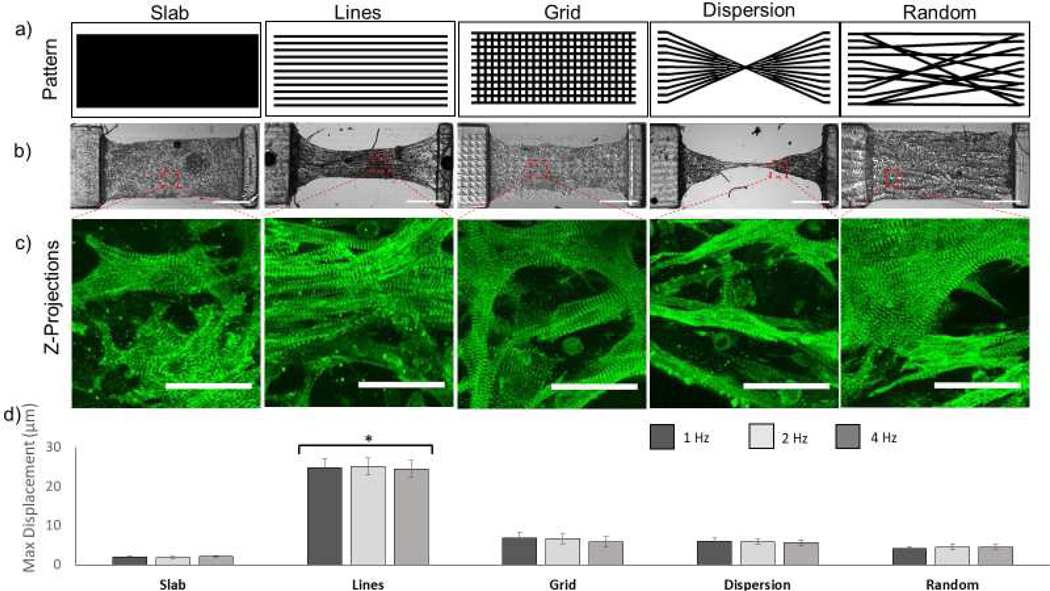
The heart possesses a complex three-dimensional (3D) laminar myofiber organization; however, because engineering physiologically relevant 3D tissues remains a technical challenge, the effects of cardiomyocyte alignment on excitation-contraction coupling, shortening and force development have not been systematically studied. Cellular shape and orientations in 3D can be controlled by engineering scaffold microstructures and encapsulating cells near these geometric cues. Here, we show that a novel method of cell encapsulation in 3D methacrylated gelatin (GelMA) scaffolds patterned via Microscale Continuous Optical Printing (μCOP) can rapidly micropattern neonatal mouse ventricular cardiomyocytes (NMVCMs) in photocrosslinkable hydrogels. Encapsulated cardiomyocytes preferentially align with the engineered microarchitecture and can display morphology and myofibril alignment phenotypic of myocardium in vivo. Utilizing the μCOP system, an asymmetric, multi-material, cantilever-based scaffold was directly printed, so that the force produced by the microtissue was transmitted onto a single deformable pillar. Aligned 3D encapsulated NMVCM scaffolds produced nearly 2 times the force compared to aligned 2D seeded samples. To further highlight the flexibility of μCOP, NMVCMs were encapsulated in several patterns to compare the effects of varying degrees of alignment on tissue displacement and synchronicity. Well aligned myofiber cultured patterns generated 4-10 times the contractile force of less anisotropically patterned constructs. Finally, normalized fluo-4 fluorescence of NMVCM-encapsulated structures showed characteristic calcium transient waveforms that increased in magnitude and rate of decline during treatment with 100 nM isoproterenol. This novel instrumented 3D cardiac microtissue serves as a physiologically relevant in vitro model system with great potential for use in cardiac disease modeling and drug screening.
心脏具有复杂的三维(3D)层状肌纤维组织;然而,由于工程学上相关的 3D 组织仍然是一个技术挑战,因此尚未系统地研究心肌细胞排列对兴奋-收缩偶联、缩短和力发展的影响。通过工程支架微结构和在这些几何线索附近封装细胞,可以控制 3D 中的细胞形状和方向。在这里,我们展示了一种在通过微尺度连续光打印(μCOP)图案化的甲基丙烯酰化明胶(GelMA)支架中进行 3D 细胞包封的新方法,该方法可以快速微图案化新生小鼠心室心肌细胞(NMVCM)在光交联水凝胶中。封装的心肌细胞优先与工程微结构对齐,并能显示出体内心肌的形态和肌原纤维排列表型。利用 μCOP 系统,直接打印了一种不对称、多材料、基于悬臂的支架,使微组织产生的力传递到单个可变形柱上。与对齐的 2D 接种样本相比,对齐的 3D 封装 NMVCM 支架产生的力几乎增加了 2 倍。为了进一步强调 μCOP 的灵活性,将 NMVCM 封装在几种图案中,以比较不同程度对齐对组织位移和同步性的影响。排列良好的肌纤维培养图案产生的收缩力是排列程度较差的图案构建体的 4-10 倍。最后,封装有 NMVCM 的结构的归一化 fluo-4 荧光显示出特征钙瞬变波形,在用 100 nM 异丙肾上腺素处理期间,钙瞬变波形的幅度和下降速率增加。这种新型仪器化的 3D 心脏微组织作为一种具有重要应用潜力的生理相关体外模型系统,可用于心脏疾病建模和药物筛选。