Rizk Hazim Mohamed, Salah Al-Deen Mohamed Sherif Mohamed, Emam Asmaa Aly
Pediatric Dentistry, Preventive & Dental Public Health Department, Faculty of Dentistry, Suez Canal University, Egypt.
Saudi Dent J. 2020 Jul;32(5):224-231. doi: 10.1016/j.sdentj.2019.09.002. Epub 2019 Sep 21.
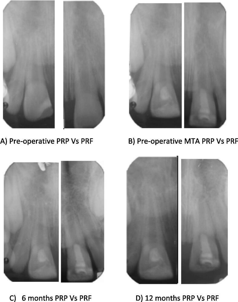
The research aims to assess the regenerative potential of Platelet Rich Plasma (PRP) versus Platelet Rich Fibrin (PRF) scaffolds in immature permanent maxillary central incisors with necrotic pulps, clinically and radiographically.
Double blinded parallel randomized controlled trial was implemented to identify the results.
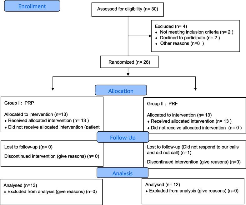
SUBJECT & METHODS: The proposed study was conducted among 30 patients with maxillary necrotic permanent immature central incisors but only 26 patients fulfilled the study requirements. Group I was treated with PRP and Group II with PRF scaffolds. Follow up has been done every 3 months for one year. Primary outcomes were measured clinically: Pain, Mobility, Swelling, and Sinus/fistula. Radiographically: increase root length and width. Secondary outcomes were clinically: Discoloration and Sensibility test. Radiographically: increase in bone density measurements and decrease in apical diameter. Standardized radiographs were collected during the follow up period, and radiographic changes were measured by using Image J software. Statistical analysis was performed on 25 patients who had completed the study.
All 25 patients' teeth were survived during the 12-month follow-up period PRP showed marginal increase in radiographic root length and width, periapical bone density and a decrease in apical diameter. No statistical significant differences were observed when it was compared with PRF. The teeth which were treated did not respond to sensibility test at the end of the study. PRF displayed statistical significant higher amount of crown discoloration when compared to PRP group
For necrotic immature teeth, revascularization using PRP is an appropriate alternative to PRF and showed excellent 12-months prognosis.
本研究旨在通过临床和影像学评估富血小板血浆(PRP)与富血小板纤维蛋白(PRF)支架在坏死牙髓的未成熟恒上颌中切牙中的再生潜力。
采用双盲平行随机对照试验来确定结果。
本研究在30例上颌坏死未成熟恒中切牙患者中进行,但只有26例患者符合研究要求。第一组接受PRP治疗,第二组接受PRF支架治疗。随访1年,每3个月进行一次。主要临床观察指标:疼痛、松动度、肿胀及窦道/瘘管。影像学观察指标:牙根长度和宽度增加。次要临床观察指标:牙齿变色和感觉测试。影像学观察指标:骨密度测量增加和根尖直径减小。在随访期间收集标准化X线片,并使用Image J软件测量影像学变化。对25例完成研究的患者进行统计分析。
在12个月的随访期内,所有25例患者的牙齿均存活。PRP组在影像学上牙根长度和宽度、根尖周骨密度有轻微增加,根尖直径减小。与PRF组相比,未观察到统计学显著差异。在研究结束时,接受治疗的牙齿对感觉测试无反应。与PRP组相比,PRF组牙齿变色在统计学上显著更严重。
对于坏死的未成熟牙齿,使用PRP进行血管再生是PRF的合适替代方法,且显示出良好的12个月预后。