• 文献检索
  • 文档翻译
  • 深度研究
  • 学术资讯
  • Suppr Zotero 插件Zotero 插件
  • 邀请有礼
  • 套餐&价格
  • 历史记录
应用&插件
Suppr Zotero 插件Zotero 插件浏览器插件Mac 客户端Windows 客户端微信小程序
定价
高级版会员购买积分包购买API积分包
服务
文献检索文档翻译深度研究API 文档MCP 服务
关于我们
关于 Suppr公司介绍联系我们用户协议隐私条款
关注我们

Suppr 超能文献

核心技术专利:CN118964589B侵权必究
粤ICP备2023148730 号-1Suppr @ 2026

文献检索

告别复杂PubMed语法,用中文像聊天一样搜索,搜遍4000万医学文献。AI智能推荐,让科研检索更轻松。

立即免费搜索

文件翻译

保留排版,准确专业,支持PDF/Word/PPT等文件格式,支持 12+语言互译。

免费翻译文档

深度研究

AI帮你快速写综述,25分钟生成高质量综述,智能提取关键信息,辅助科研写作。

立即免费体验

低剂量计算机断层扫描筛查中同步自动冠状动脉钙化评分的未来

The Future of Concurrent Automated Coronary Artery Calcium Scoring on Screening Low-Dose Computed Tomography.

作者信息

Waltz Jeffrey, Kocher Madison, Kahn Jacob, Dirr McKenzie, Burt Jeremy R

机构信息

Diagnostic Radiology, Medical University of South Carolina, Charleston, USA.

Radiology, Medical University of South Carolina, Charleston, USA.

出版信息

Cureus. 2020 Jun 12;12(6):e8574. doi: 10.7759/cureus.8574.

DOI:10.7759/cureus.8574
PMID:32670710
原文链接:https://pmc.ncbi.nlm.nih.gov/articles/PMC7358941/
Abstract

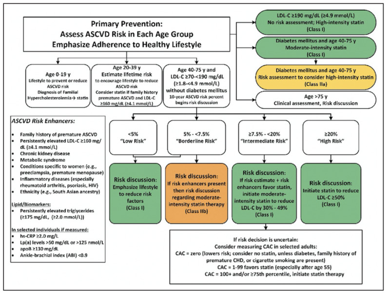
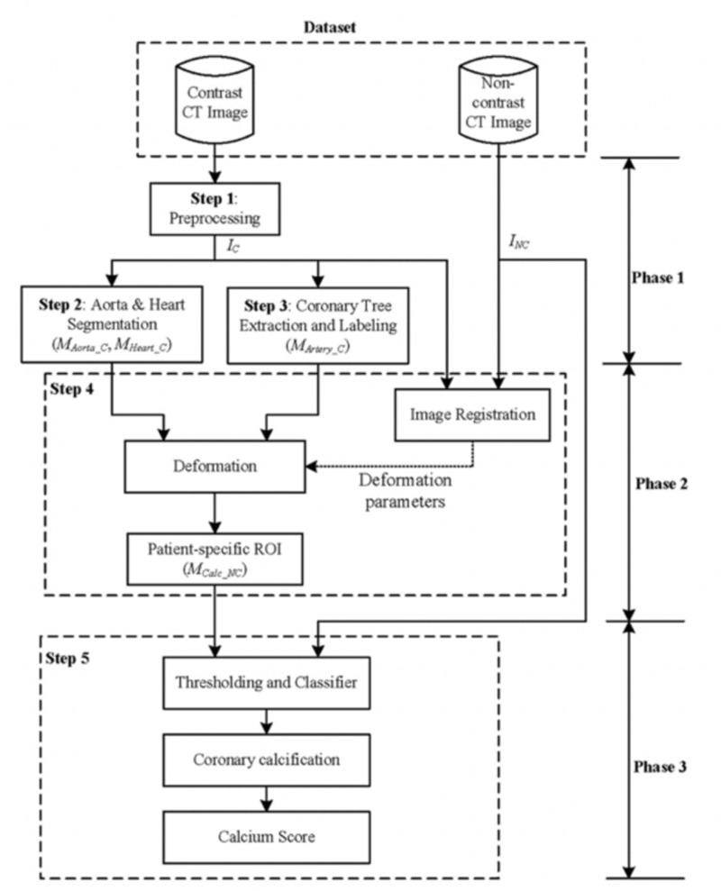
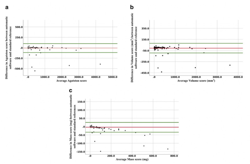
Low-dose computed tomography (LDCT) has been extensively validated for lung cancer screening in selected patient populations. Additionally, the use of gated cardiac CT to assess coronary artery calcium (CAC) burden has been validated to determine a patient's risk for major cardiovascular adverse events. This is typically performed by calculating an Agatston score based on density and overall burden of calcified plaque within the coronary arteries. Patients that qualify for LDCT for lung cancer screening commonly share major risk factors for coronary artery disease and would frequently benefit from an additional gated cardiac CT for the assessment of CAC. Given the widespread use of LDCT for lung cancer screening, we evaluated current literature regarding the use of non-gated chest CT, specifically LDCT, for the detection and grading of coronary artery calcifications. Additionally, given the evolving and increasing use of artificial intelligence (AI) in the interpretation of radiologic studies, current literature for the use of AI in CAC assessment was reviewed.  We reviewed primary scientific literature dating up to April 2020 using Pubmed and Google Scholar, with the search terms low dose CT, lung cancer screening, coronary artery calcium, EKG/cardiac gated CT, deep learning, machine learning, and AI. These publications were then independently evaluated by each member of our team. Overall, there was a consensus within these papers that LDCT for lung cancer screening plays a role in the evaluation of CAC. Most studies note the inherent problems with the evaluation of the density of coronary calcifications on LDCT to give an accurate numeric calcium or Agatston score. The current method of evaluating CAC on LDCT involves using a qualitative categorical system (none, mild, moderate, or severe). When performed by cardiac imaging experts, this method broadly correlates with traditional CAC score groups (0, 1 to 100, 101 to 400, and > 400). Furthermore, given the high sensitivity of a properly protocolled LDCT for coronary calcium, a negative study for CAC precludes the need for a dedicated gated CT assessment. However, qualitative methods are not as accurate or reproducible when performed by general radiologists. The implementation of AI in the LDCT screening process has the potential to give a quantifiable and reproducible numeric value to the calcium score, based on whole heart volume scoring of calcium. This more closely aligns with the Agatston score and serves as a better guide for treatment and risk assessment using current guidelines. We conclude that CAC should be assessed on all LDCT performed for lung cancer screening and that a qualitative categorical scoring system should be provided in the impression for each patient. Early studies involving AI for the assessment of CAC are promising, but more extensive studies are needed before a final recommendation for its use can be given. The implementation of an accurate, automated AI CAC assessment tool would improve radiologist compliance and ease of overall workflow. Ultimately, the potential end result would be improved turnaround time, better patient outcomes, and reduced healthcare costs by maximizing preventative care in this high-risk population.

摘要

低剂量计算机断层扫描(LDCT)已在特定患者群体中广泛用于肺癌筛查。此外,使用门控心脏CT评估冠状动脉钙化(CAC)负荷已被证实可用于确定患者发生重大心血管不良事件的风险。这通常通过基于冠状动脉内钙化斑块的密度和总体负荷计算阿加斯顿评分来进行。符合LDCT肺癌筛查条件的患者通常具有冠状动脉疾病的主要危险因素,并且经常会受益于额外的门控心脏CT以评估CAC。鉴于LDCT在肺癌筛查中的广泛应用,我们评估了有关使用非门控胸部CT(特别是LDCT)进行冠状动脉钙化检测和分级的现有文献。此外,鉴于人工智能(AI)在放射学研究解读中的应用不断发展且日益增多,我们还回顾了有关AI在CAC评估中应用的现有文献。我们使用PubMed和谷歌学术搜索了截至2020年4月的主要科学文献,搜索词包括低剂量CT、肺癌筛查、冠状动脉钙化、心电图/心脏门控CT、深度学习、机器学习和AI。然后我们团队的每个成员对这些出版物进行了独立评估。总体而言,这些论文达成的共识是,用于肺癌筛查的LDCT在CAC评估中发挥作用。大多数研究指出,在LDCT上评估冠状动脉钙化密度以给出准确的数字钙或阿加斯顿评分存在固有问题。目前在LDCT上评估CAC的方法涉及使用定性分类系统(无、轻度、中度或重度)。由心脏影像专家进行时,这种方法与传统的CAC评分组(0、1至100、101至400以及>400)大致相关。此外,鉴于适当方案的LDCT对冠状动脉钙化的高敏感性,CAC的阴性研究排除了进行专门门控CT评估的必要性。然而,当由普通放射科医生进行时,定性方法的准确性和可重复性较差。在LDCT筛查过程中实施AI有可能基于对心脏整体体积的钙评分给出可量化且可重复的钙数值。这与阿加斯顿评分更接近,并且作为使用当前指南进行治疗和风险评估的更好指导。我们得出结论,对于所有用于肺癌筛查的LDCT都应评估CAC,并且应在每份患者的影像报告中提供定性分类评分系统。涉及AI评估CAC的早期研究很有前景,但在给出最终使用建议之前还需要更广泛的研究。实施准确、自动化的AI CAC评估工具将提高放射科医生的依从性并简化整体工作流程。最终,潜在的最终结果将是通过在这个高风险人群中最大化预防保健来缩短周转时间、改善患者预后并降低医疗成本。

https://cdn.ncbi.nlm.nih.gov/pmc/blobs/45b9/7358941/43a1da0d28eb/cureus-0012-00000008574-i05.jpg
https://cdn.ncbi.nlm.nih.gov/pmc/blobs/45b9/7358941/b67d83eef8eb/cureus-0012-00000008574-i01.jpg
https://cdn.ncbi.nlm.nih.gov/pmc/blobs/45b9/7358941/1e7f1b664f2f/cureus-0012-00000008574-i02.jpg
https://cdn.ncbi.nlm.nih.gov/pmc/blobs/45b9/7358941/4a055096d511/cureus-0012-00000008574-i03.jpg
https://cdn.ncbi.nlm.nih.gov/pmc/blobs/45b9/7358941/e39aef431000/cureus-0012-00000008574-i04.jpg
https://cdn.ncbi.nlm.nih.gov/pmc/blobs/45b9/7358941/43a1da0d28eb/cureus-0012-00000008574-i05.jpg

相似文献

1
The Future of Concurrent Automated Coronary Artery Calcium Scoring on Screening Low-Dose Computed Tomography.低剂量计算机断层扫描筛查中同步自动冠状动脉钙化评分的未来
Cureus. 2020 Jun 12;12(6):e8574. doi: 10.7759/cureus.8574.
2
Integrating cardiovascular risk assessment into mobile low-dose CT lung screenings in rural Appalachia: A comprehensive analysis of the relationship between lung cancer risk, coronary artery calcium burden, and cardiovascular risk reduction strategies.将心血管风险评估纳入阿巴拉契亚农村地区的移动低剂量CT肺癌筛查:对肺癌风险、冠状动脉钙化负荷和心血管风险降低策略之间关系的综合分析。
Am J Prev Cardiol. 2024 Aug 11;19:100719. doi: 10.1016/j.ajpc.2024.100719. eCollection 2024 Sep.
3
Coronary Artery Calcification on Low-Dose Lung Cancer Screening CT in South Korea: Visual and Artificial Intelligence-Based Assessment and Association With Cardiovascular Events.韩国低剂量肺癌筛查 CT 冠状动脉钙化:基于视觉和人工智能的评估及其与心血管事件的关系。
AJR Am J Roentgenol. 2024 May;222(5):e2430852. doi: 10.2214/AJR.24.30852. Epub 2024 May 29.
4
Reliability analysis of visual ranking of coronary artery calcification on low-dose CT of the thorax for lung cancer screening: comparison with ECG-gated calcium scoring CT.用于肺癌筛查的胸部低剂量CT上冠状动脉钙化视觉分级的可靠性分析:与心电图门控钙评分CT的比较
Int J Cardiovasc Imaging. 2014 Dec;30 Suppl 2:81-7. doi: 10.1007/s10554-014-0507-8. Epub 2014 Aug 2.
5
Fully automatic coronary calcium scoring in non-ECG-gated low-dose chest CT: comparison with ECG-gated cardiac CT.非心电门控低剂量胸部CT中的全自动冠状动脉钙化评分:与心电门控心脏CT的比较
Eur Radiol. 2023 Feb;33(2):1254-1265. doi: 10.1007/s00330-022-09117-3. Epub 2022 Sep 13.
6
Automated total and vessel-specific coronary artery calcium (CAC) quantification on chest CT: direct comparison with CAC scoring on non-contrast cardiac CT.基于胸部 CT 的全自动冠状动脉钙化(CAC)定量分析:与非增强心脏 CT 的 CAC 评分的直接比较。
BMC Med Imaging. 2022 Oct 14;22(1):177. doi: 10.1186/s12880-022-00907-1.
7
Evaluation of fully automated commercial software for Agatston calcium scoring on non-ECG-gated low-dose chest CT with different slice thickness.在不同层厚的非心电图门控低剂量胸部CT上,对用于阿加斯顿钙评分的全自动商业软件进行评估。
Eur Radiol. 2023 Mar;33(3):1973-1981. doi: 10.1007/s00330-022-09143-1. Epub 2022 Sep 24.
8
Evaluation of Clinical Applicability of Coronary Artery Calcium Assessment on Non-Gated Chest Computed Tomography, Compared With the Classic Agatston Score on Cardiac Computed Tomography.非门控胸部计算机断层扫描冠状动脉钙化评估的临床适用性评估,与心脏计算机断层扫描上的经典阿加特斯顿评分对比
Am J Cardiol. 2023 Dec 1;208:92-100. doi: 10.1016/j.amjcard.2023.08.186. Epub 2023 Oct 10.
9
The prognostic value of visual and automatic coronary calcium scoring from low-dose computed tomography-[15O]-water positron emission tomography.低剂量计算机断层扫描-[15O]-水正电子发射断层扫描的视觉和自动冠状动脉钙评分的预后价值。
Eur Heart J Cardiovasc Imaging. 2024 Aug 26;25(9):1186-1196. doi: 10.1093/ehjci/jeae081.
10
Estimation of cardiovascular risk on routine chest CT: Ordinal coronary artery calcium scoring as an accurate predictor of Agatston score ranges.常规胸部CT评估心血管风险:序贯冠状动脉钙化积分作为阿加斯顿积分范围的准确预测指标。
J Cardiovasc Comput Tomogr. 2017 Jan-Feb;11(1):8-15. doi: 10.1016/j.jcct.2016.10.001. Epub 2016 Oct 5.

引用本文的文献

1
Coronary artery calcification score as a prognostic factor in younger patients with multiple myeloma undergoing autologous stem cell therapy.冠状动脉钙化评分作为接受自体干细胞治疗的年轻多发性骨髓瘤患者的预后因素。
Cardiooncology. 2025 Apr 23;11(1):38. doi: 10.1186/s40959-025-00338-1.
2
A Thorough Review of the Clinical Applications of Artificial Intelligence in Lung Cancer.人工智能在肺癌临床应用的全面综述
Cancers (Basel). 2025 Mar 4;17(5):882. doi: 10.3390/cancers17050882.
3
The Clear Value of Coronary Artery Calcification Evaluation on Non-Gated Chest Computed Tomography for Cardiac Risk Stratification.

本文引用的文献

1
The American Cancer Society's Facts & Figures: 2020 Edition.美国癌症协会《2020年事实与数据》版
J Adv Pract Oncol. 2020 Mar;11(2):135-136. doi: 10.6004/jadpro.2020.11.2.1. Epub 2020 Mar 1.
2
Hybrid Neural Networks for Mortality Prediction from LDCT Images.用于从低剂量CT图像预测死亡率的混合神经网络。
Annu Int Conf IEEE Eng Med Biol Soc. 2019 Jul;2019:6243-6246. doi: 10.1109/EMBC.2019.8857180.
3
Cancer statistics, 2020.癌症统计数据,2020 年。
非门控胸部计算机断层扫描评估冠状动脉钙化对心脏风险分层的明确价值
Cardiol Ther. 2024 Mar;13(1):69-87. doi: 10.1007/s40119-024-00354-9. Epub 2024 Feb 13.
4
Role and progress of artificial intelligence in radiodiagnosing vascular calcification: a narrative review.人工智能在血管钙化放射诊断中的作用与进展:一篇叙述性综述
Ann Transl Med. 2023 Jan 31;11(2):131. doi: 10.21037/atm-22-6333. Epub 2023 Jan 13.
5
Reporting Coronary Artery Calcium on Low-Dose Computed Tomography Impacts Statin Management in a Lung Cancer Screening Population.低剂量计算机断层扫描报告冠状动脉钙化对肺癌筛查人群的他汀类药物管理有影响。
Fed Pract. 2022 Sep;39(9):382-388. doi: 10.12788/fp.0318. Epub 2022 Sep 14.
6
The Potential Role of Artificial Intelligence in Lung Cancer Screening Using Low-Dose Computed Tomography.人工智能在低剂量计算机断层扫描肺癌筛查中的潜在作用
Diagnostics (Basel). 2022 Oct 8;12(10):2435. doi: 10.3390/diagnostics12102435.
7
Evaluation of fully automated commercial software for Agatston calcium scoring on non-ECG-gated low-dose chest CT with different slice thickness.在不同层厚的非心电图门控低剂量胸部CT上,对用于阿加斯顿钙评分的全自动商业软件进行评估。
Eur Radiol. 2023 Mar;33(3):1973-1981. doi: 10.1007/s00330-022-09143-1. Epub 2022 Sep 24.
8
Design and Rationale of the HANSE Study: A Holistic German Lung Cancer Screening Trial Using Low-Dose Computed Tomography.HANSE 研究的设计和原理:使用低剂量计算机断层扫描的整体德国肺癌筛查试验。
Rofo. 2022 Dec;194(12):1333-1345. doi: 10.1055/a-1853-8291. Epub 2022 Aug 2.
9
Artificial intelligence and imaging: Opportunities in cardio-oncology.人工智能与成像:心脏肿瘤学中的机遇。
Am Heart J Plus. 2022 Mar;15. doi: 10.1016/j.ahjo.2022.100126. Epub 2022 Apr 6.
10
Classification of moving coronary calcified plaques based on motion artifacts using convolutional neural networks: a robotic simulating study on influential factors.基于运动伪影的运动性冠状动脉钙化斑块分类:机器人模拟研究影响因素。
BMC Med Imaging. 2021 Oct 19;21(1):151. doi: 10.1186/s12880-021-00680-7.
CA Cancer J Clin. 2020 Jan;70(1):7-30. doi: 10.3322/caac.21590. Epub 2020 Jan 8.
4
Evaluation of an AI-based, automatic coronary artery calcium scoring software.基于人工智能的自动冠状动脉钙评分软件的评估。
Eur Radiol. 2020 Mar;30(3):1671-1678. doi: 10.1007/s00330-019-06489-x. Epub 2019 Nov 14.
5
2019 ACC/AHA Guideline on the Primary Prevention of Cardiovascular Disease: Executive Summary: A Report of the American College of Cardiology/American Heart Association Task Force on Clinical Practice Guidelines.2019 ACC/AHA 心血管疾病一级预防指南:执行摘要:美国心脏病学会/美国心脏协会临床实践指南工作组的报告。
J Am Coll Cardiol. 2019 Sep 10;74(10):1376-1414. doi: 10.1016/j.jacc.2019.03.009. Epub 2019 Mar 17.
6
Comparison of Nongated Chest CT and Dedicated Calcium Scoring CT for Coronary Calcium Quantification Using a 256-Dector Row CT Scanner.使用 256 排 CT 扫描仪对非门控胸部 CT 与专用钙评分 CT 进行冠状动脉钙化定量的比较。
Acad Radiol. 2019 Oct;26(10):e267-e274. doi: 10.1016/j.acra.2018.12.005. Epub 2019 Jan 23.
7
2018 AHA/ACC/AACVPR/AAPA/ABC/ACPM/ADA/AGS/APhA/ASPC/NLA/PCNA Guideline on the Management of Blood Cholesterol: A Report of the American College of Cardiology/American Heart Association Task Force on Clinical Practice Guidelines.2018 年美国心脏病学会/美国心脏协会/美国心血管血管造影和介入学会/美国预防、物理治疗和康复医师学会/美国糖尿病协会/美国老年学会/美国药学会/美国医师协会/美国生理学会/北美介入放射学会/美国国家脂质协会/美国临床内分泌医师协会胆固醇管理指南:美国心脏病学会/美国心脏协会临床实践指南工作组的报告。
Circulation. 2019 Jun 18;139(25):e1082-e1143. doi: 10.1161/CIR.0000000000000625. Epub 2018 Nov 10.
8
Lung Cancer Screening Inconsistent With U.S. Preventive Services Task Force Recommendations.肺癌筛查不符合美国预防服务工作组的建议。
Am J Prev Med. 2019 Jan;56(1):66-73. doi: 10.1016/j.amepre.2018.07.030. Epub 2018 Nov 19.
9
Optimized categorization algorithm of coronary artery calcification score on non-gated chest low-dose CT screening using iterative model reconstruction technique.基于迭代模型重建技术的非门控胸部低剂量CT筛查冠状动脉钙化积分优化分类算法
Clin Imaging. 2018 Nov-Dec;52:287-291. doi: 10.1016/j.clinimag.2018.08.015. Epub 2018 Aug 29.
10
Automated Agatston Score Computation in non-ECG Gated CT Scans Using Deep Learning.使用深度学习在非心电图门控CT扫描中自动计算阿加西分数
Proc SPIE Int Soc Opt Eng. 2018 Feb;10574. doi: 10.1117/12.2293681. Epub 2018 Mar 2.