Liu Bingchuan, Zhao Yanran, Zhu Tengjiao, Gao Shan, Ye Kaifeng, Zhou Fang, Qiu Dong, Wang Xing, Tian Yun, Qu Xiaozhong
Department of Orthopaedics, Peking University Third Hospital, Beijing, China.
Engineering Research Center of Bone and Joint Precision Medicine, Ministry of Education, Peking University Third Hospital, Beijing, China.
Front Bioeng Biotechnol. 2020 Jul 2;8:752. doi: 10.3389/fbioe.2020.00752. eCollection 2020.
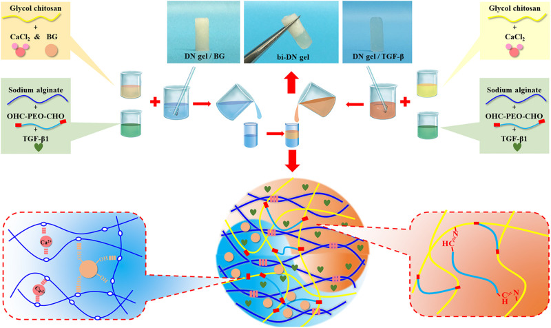
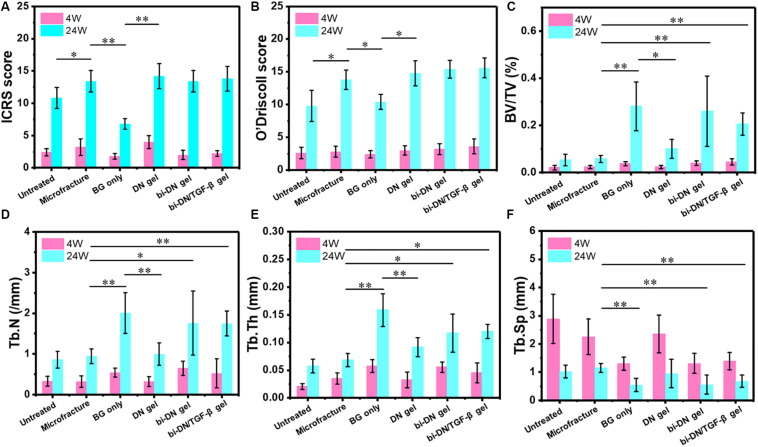
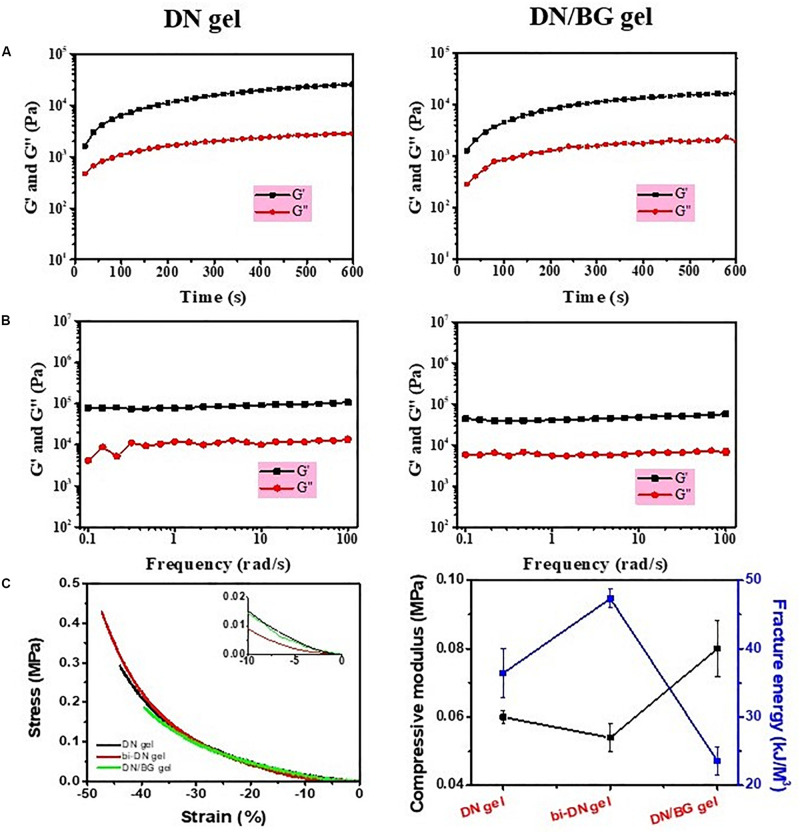
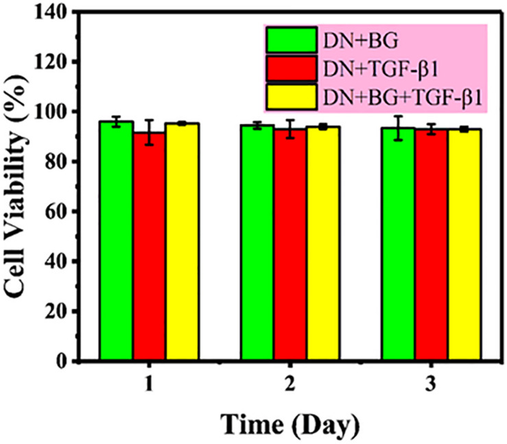
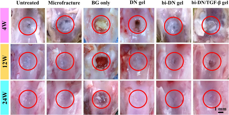
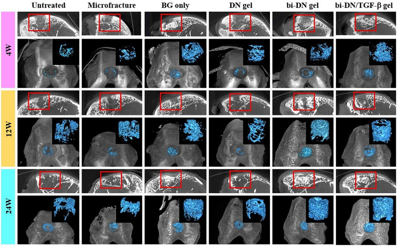
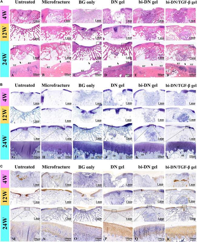
Periarticular injury usually causes the defects of superficial cartilage and the underlying subchondral bone. Although some efficacious outcomes have been achieved by the existing therapeutic methods both in clinics and research, like symptomatic treatment, microfracture surgery, and tissue engineering technology, they still present specific disadvantages and complications. To improve this situation, we designed a biphasic (bi-) scaffold aiming to repair the structure of cartilage and subchondral bone synchronously. The scaffold consisted of a superior double-network (DN) hydrogel layer and a lower bioactive glass (BG) reinforced hydrogel layer, and the DN hydrogel included glycol chitosan (GC) and dibenzaldhyde functionalized poly(ethylene oxide) network, and sodium alginate (Alg) and calcium chloride (CaCl) network. To investigate its effectiveness, we applied this biphasic scaffold to repair osteochondral full-thickness defects in rabbit models. We set up six observation groups in total, including Untreated group, Microfracture group, BG only group, DN gel group, bi-DN gel group, and bi-DN/TGF-β gel group. With a follow-up period of 24 weeks, we evaluated the treatment effects by gross observation, micro-CT scan and histological staining. Besides, we further fulfilled the quantitative analysis of the data from ICRS score, O'Driscoll score and micro-CT parameters. The results revealed that neat GC/Alg DN hydrogel scaffold was only conductive to promoting cartilage regeneration and neat BG scaffold merely showed the excellent ability to reconstruct subchondral bone. While the biphasic scaffold performed better in repairing osteochondral defect synchronously, exhibiting more well-integrated cartilage-like tissue with positive staining of toluidine blue and col II immunohistochemistry, and more dense trabecular bone connecting closely with the surrounding host bone. Therefore, this method possessed the clinical application potential in treating articular injury, osteochondral degeneration, osteochondral necrosis, and sclerosis.
关节周围损伤通常会导致表层软骨和其下方的软骨下骨出现缺损。尽管现有治疗方法在临床和研究中都取得了一些有效的成果,如对症治疗、微骨折手术和组织工程技术,但它们仍然存在特定的缺点和并发症。为改善这种情况,我们设计了一种双相支架,旨在同步修复软骨和软骨下骨的结构。该支架由上层的双网络(DN)水凝胶层和下层的生物活性玻璃(BG)增强水凝胶层组成,DN水凝胶包括乙二醇壳聚糖(GC)和二苯甲醛功能化聚环氧乙烷网络,以及海藻酸钠(Alg)和氯化钙(CaCl)网络。为研究其有效性,我们将这种双相支架应用于兔模型的骨软骨全层缺损修复。我们总共设置了六个观察组,包括未治疗组、微骨折组、仅BG组、DN凝胶组、双相DN凝胶组和双相DN/TGF-β凝胶组。随访24周后,我们通过大体观察、显微CT扫描和组织学染色评估治疗效果。此外,我们还对国际软骨修复协会(ICRS)评分、奥德里斯科尔(O'Driscoll)评分和显微CT参数的数据进行了进一步的定量分析。结果表明,单纯的GC/Alg DN水凝胶支架仅有助于促进软骨再生,而单纯的BG支架仅显示出重建软骨下骨的出色能力。而双相支架在同步修复骨软骨缺损方面表现更好,表现出更多整合良好的软骨样组织,甲苯胺蓝和II型胶原免疫组化染色呈阳性,以及更致密的小梁骨与周围宿主骨紧密相连。因此,这种方法在治疗关节损伤、骨软骨退变、骨软骨坏死和硬化方面具有临床应用潜力。