Kubelick Kelsey P, Emelianov Stanislav Y
Georgia Institute of Technology, Emory University School of Medicine, Wallace H. Coulter Department of Biomedical Engineering, Atlanta, Georgia, United States.
Georgia Institute of Technology, School of Electrical and Computer Engineering, Atlanta, Georgia, United States.
Neurophotonics. 2020 Jul;7(3):030501. doi: 10.1117/1.NPh.7.3.030501. Epub 2020 Jul 25.
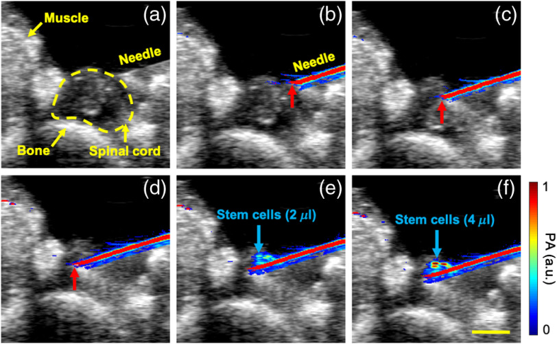
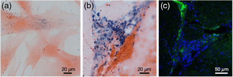
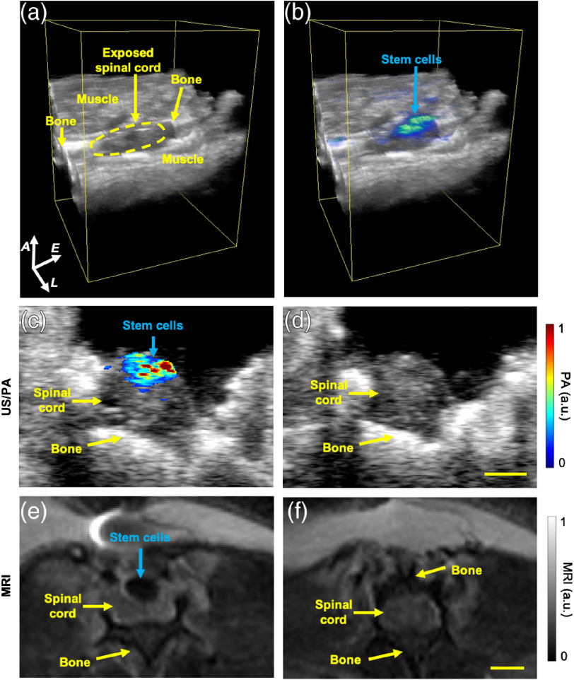
Stem cell therapies are of interest for treating a variety of neurodegenerative diseases and injuries of the spinal cord. However, the lack of techniques for longitudinal monitoring of stem cell therapy progression is inhibiting clinical translation. The goal of this study is to demonstrate an intraoperative imaging approach to guide stem cell injection to the spinal cord . Results may ultimately support the development of an imaging tool that spans intra- or postoperative environments to guide therapy throughout treatment. Stem cells were labeled with Prussian blue nanocubes (PBNCs) to facilitate combined ultrasound and photoacoustic (US/PA) imaging to visualize stem cell injection and delivery to the spinal cord . US/PA results were confirmed by magnetic resonance imaging (MRI) and histology. Real-time intraoperative US/PA image-guided injection of PBNC-labeled stem cells and three-dimensional volumetric images of injection provided feedback necessary for successful delivery of therapeutics into the spinal cord. Postoperative MRI confirmed delivery of PBNC-labeled stem cells. The nanoparticle-augmented US/PA approach successfully detected injection and delivery of stem cells into the spinal cord, confirmed by MRI. Our work demonstrated feasibility, which is a critical step toward the development of a US/PA/MRI platform to monitor regenerative spinal cord therapies.
干细胞疗法对于治疗多种神经退行性疾病和脊髓损伤具有重要意义。然而,缺乏对干细胞治疗进展进行纵向监测的技术阻碍了其临床转化。本研究的目的是展示一种术中成像方法,以指导干细胞向脊髓的注射。研究结果最终可能支持开发一种跨越术中或术后环境的成像工具,以在整个治疗过程中指导治疗。用普鲁士蓝纳米立方体(PBNCs)标记干细胞,以促进超声和光声(US/PA)联合成像,从而可视化干细胞向脊髓的注射和递送。US/PA结果通过磁共振成像(MRI)和组织学得到证实。实时术中US/PA图像引导下注射PBNC标记的干细胞以及注射的三维体积图像为将治疗药物成功递送至脊髓提供了必要的反馈。术后MRI证实了PBNC标记的干细胞的递送。纳米颗粒增强的US/PA方法成功检测到干细胞向脊髓的注射和递送,并得到MRI的证实。我们的工作证明了其可行性,这是朝着开发用于监测脊髓再生治疗的US/PA/MRI平台迈出的关键一步。