Department of Neurosciences-Laboratory of Neuropathology, Campus Gasthuisberg (O&N4), KU-Leuven, Herestraat 49, 3000, Leuven, Belgium.
Department of Pathology, UZ-Leuven, Leuven, Belgium.
Acta Neuropathol. 2018 Oct;136(4):557-567. doi: 10.1007/s00401-018-1897-9. Epub 2018 Aug 19.
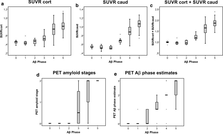
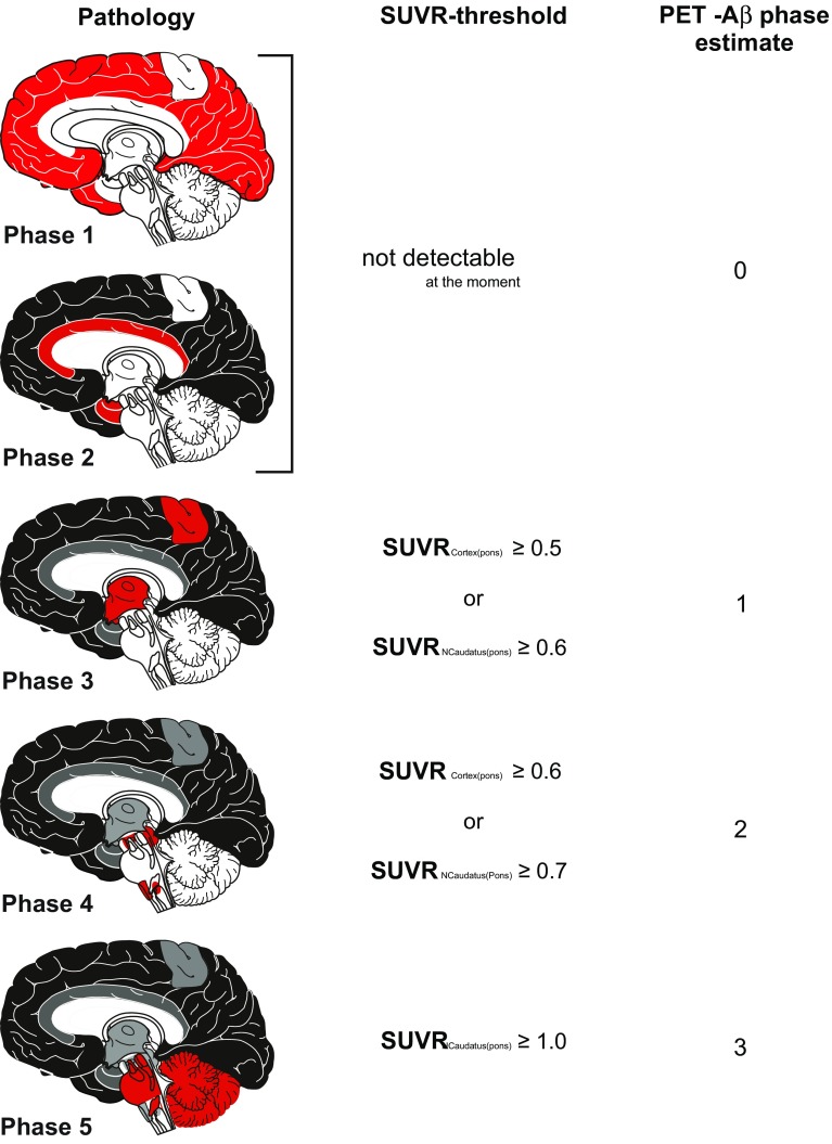
The deposition of the amyloid β-protein (Aβ) in senile plaques is one of the histopathological hallmarks of Alzheimer's disease (AD). Aβ-plaques arise first in neocortical areas and, then, expand into further brain regions in a process described by 5 phases. Since it is possible to identify amyloid pathology with radioactive-labeled tracers by positron emission tomography (PET) the question arises whether it is possible to distinguish the neuropathological Aβ-phases with amyloid PET imaging. To address this question we reassessed 97 cases of the end-of-life study cohort of the phase 3 [F]flutemetamol trial (ClinicalTrials.gov identifiers NCT01165554, and NCT02090855) by combining the standardized uptake value ratios (SUVRs) with pons as reference region for cortical and caudate nucleus-related [F]flutemetamol-retention. We tested them for their prediction of the neuropathological pattern found at autopsy. By defining threshold levels for cortical and caudate nucleus SUVRs we could distinguish different levels of [F]flutemetamol uptake termed PET-Aβ phase estimates. When comparing these PET-Aβ phase estimates with the neuropathological Aβ-phases we found that PET-Aβ phase estimate 0 corresponded with Aβ-phases 0-2, 1 with Aβ-phase 3, 2 with Aβ-phase 4, and 3 with Aβ-phase 5. Classification using the PET-Aβ phase estimates predicted the correct Aβ-phase in 72.16% of the cases studied here. Bootstrap analysis was used to confirm the robustness of the estimates around this association. When allowing a range of ± 1 phase for a given Aβ-phase correct classification was given in 96.91% of the cases. In doing so, we provide a novel method to convert SUVR-levels into PET-Aβ phase estimates that can be easily translated into neuropathological phases of Aβ-deposition. This method allows direct conclusions about the pathological distribution of amyloid plaques (Aβ-phases) in vivo. Accordingly, this method may be ideally suited to detect early preclinical AD-patients, to follow them with disease progression, and to provide a more precise prognosis for them based on the knowledge about the underlying pathological phase of the disease.
β淀粉样蛋白(Aβ)在老年斑中的沉积是阿尔茨海默病(AD)的组织病理学标志之一。Aβ 斑块首先出现在新皮质区域,然后在 5 个阶段的过程中扩展到大脑的其他区域。由于正电子发射断层扫描(PET)可以用放射性标记示踪剂识别淀粉样蛋白病理学,因此出现了一个问题,即用淀粉样蛋白 PET 成像是否可以区分神经病理学 Aβ 阶段。为了解决这个问题,我们通过将皮质和尾状核的标准化摄取值比值(SUVR)与桥脑作为参考区域相结合,重新评估了 3 期[F]flutemetamol 试验(ClinicalTrials.gov 标识符 NCT01165554 和 NCT02090855)的临终研究队列中的 97 例病例。我们测试了它们对尸检中发现的神经病理学模式的预测能力。通过定义皮质和尾状核 SUVR 的阈值水平,我们可以区分不同水平的[F]flutemetamol 摄取,称为 PET-Aβ 阶段估计。当将这些 PET-Aβ 阶段估计与神经病理学 Aβ 阶段进行比较时,我们发现 PET-Aβ 阶段估计 0 与 Aβ 阶段 0-2 相对应,1 与 Aβ 阶段 3 相对应,2 与 Aβ 阶段 4 相对应,3 与 Aβ 阶段 5 相对应。使用 PET-Aβ 阶段估计进行分类,可在研究的 72.16%的病例中正确预测 Aβ 阶段。使用 bootstrap 分析证实了这种关联的估计值的稳健性。当允许给定 Aβ 阶段的±1 个阶段时,96.91%的病例可以得到正确分类。通过这样做,我们提供了一种将 SUVR 水平转换为 PET-Aβ 阶段估计的新方法,该方法可以很容易地转化为 Aβ 沉积的神经病理学阶段。这种方法可以直接得出体内淀粉样斑块(Aβ 阶段)的病理分布的结论。因此,该方法可能非常适合于检测早期的临床前 AD 患者,随着疾病的进展对他们进行跟踪,并根据对疾病潜在病理阶段的了解为他们提供更准确的预后。