Prange Lyndsey, Pratt Milton, Herman Kristin, Schiffmann Raphael, Mueller David M, McLean Melissa, Mendez Mary Moya, Walley Nicole, Heinzen Erin L, Goldstein David, Shashi Vandana, Hunanyan Arsen, Pagadala Vijay, Mikati Mohamad A
Duke University (L.P., M.P., M.M.M., N.W., V.S., A.H., M.A.M.), Durham, NC; UC Davis Health (K.H.), Sacramento; Baylor Scott & White Health (R.S.), Dallas, TX; Rosalind Franklin University of Medicine and Science (D.M.M.), Chicago, IL; University of North Carolina at Chapel Hill (E.L.H.); Columbia University (D.G.), New York City, NY; and Glycan Therapeutics, LLC (V.P.), Chapel Hill, NC.
Neurol Genet. 2020 Aug 4;6(5):e466. doi: 10.1212/NXG.0000000000000466. eCollection 2020 Oct.
To describe a phenotype caused by mutations, which manifests as dystonia, dysmorphism of the face, encephalopathy with developmental delay, brain MRI abnormalities always including cerebellar hypoplasia, no hemiplegia (Ø) (D-DEMØ), and neonatal onset.
Review and analysis of clinical and genetic data.
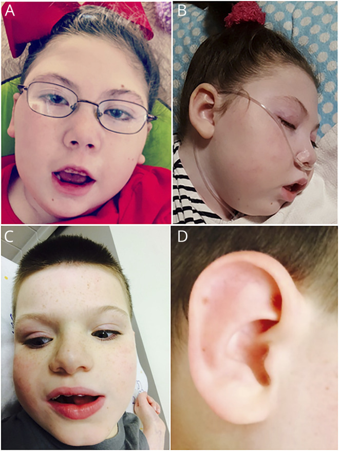
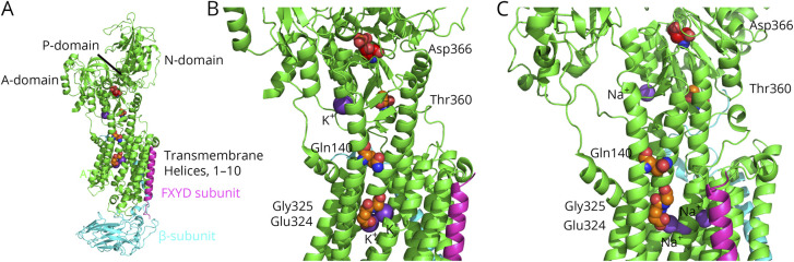
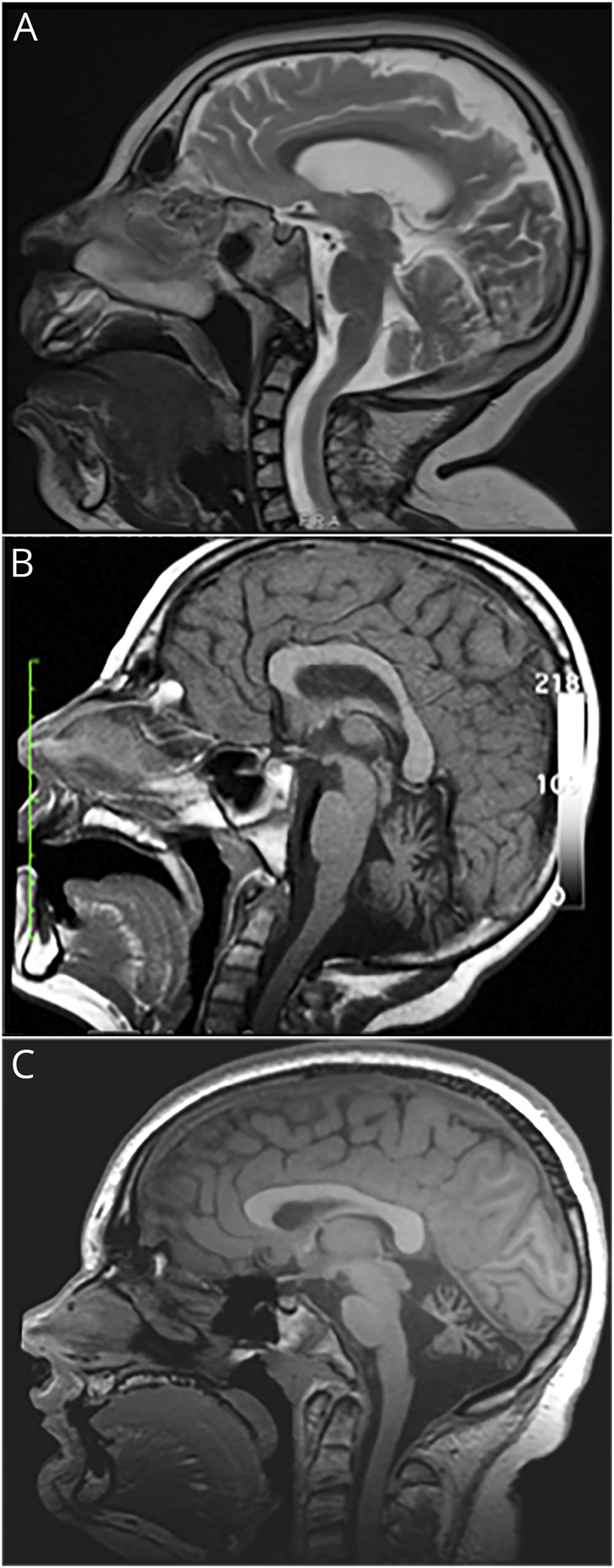
Patients shared the above traits and had whole-exome sequencing that showed de novo variants of the gene, predicted to be disease causing and occurring in regions of the protein critical for pump function. Patient 1 (c.1079C>G, p.Thr360Arg), an 8-year-old girl, presented on day 1 of life with episodic dystonia, complex partial seizures, and facial dysmorphism. MRI of the brain revealed cerebellar hypoplasia. Patient 2 (c.420G>T, p.Gln140His), an 18-year-old man, presented on day 1 of life with hypotonia, tremor, and facial dysmorphism. He later developed dystonia. MRI of the brain revealed cerebellar hypoplasia and, later, further cerebellar volume loss (atrophy). Patient 3 (c.974G>A, Gly325Asp), a 13-year-old girl, presented on day 1 of life with tremor, episodic dystonia, and facial dysmorphism. MRI of the brain showed severe cerebellar hypoplasia. Patient 4 (c.971A>G, p.Glu324Gly), a 14-year-old boy, presented on day 1 of life with tremor, hypotonia, dystonia, nystagmus, facial dysmorphism, and later seizures. MRI of the brain revealed moderate cerebellar hypoplasia.
D-DEMØ represents an -related phenotype, the observation of which should trigger investigation for mutations. Our findings, and the presence of multiple distinct -related phenotypes, support the possibility that there are differences in the underlying mechanisms.
描述一种由突变引起的表型,其表现为肌张力障碍、面部畸形、伴有发育迟缓的脑病、脑部MRI异常(总是包括小脑发育不全)、无偏瘫(Ø)(D-DEMØ)以及新生儿期起病。
回顾和分析临床及基因数据。
患者具有上述特征,且全外显子测序显示该基因的新生变异,预计为致病变异且发生在对泵功能至关重要的蛋白质区域。患者1(c.1079C>G,p.Thr360Arg),一名8岁女孩,出生第1天出现发作性肌张力障碍、复杂部分性发作和面部畸形。脑部MRI显示小脑发育不全。患者2(c.420G>T,p.Gln140His),一名18岁男性,出生第1天出现肌张力低下、震颤和面部畸形。他后来发展为肌张力障碍。脑部MRI显示小脑发育不全,随后小脑体积进一步减小(萎缩)。患者3(c.974G>A,Gly325Asp),一名13岁女孩,出生第1天出现震颤、发作性肌张力障碍和面部畸形。脑部MRI显示严重小脑发育不全。患者4(c.971A>G,p.Glu324Gly),一名14岁男孩,出生第1天出现震颤、肌张力低下、肌张力障碍、眼球震颤、面部畸形,后来出现癫痫发作。脑部MRI显示中度小脑发育不全。
D-DEMØ代表一种与[基因名称]相关的表型,对其观察应引发对[基因名称]突变的调查。我们的发现以及多种不同的与[基因名称]相关表型的存在,支持了潜在机制存在差异的可能性。