Gestermann Nicolas, Saugy Damien, Martignier Christophe, Tillé Laure, Fuertes Marraco Silvia A, Zettl Markus, Tirapu Iñigo, Speiser Daniel E, Verdeil Grégory
Department of Oncology UNIL CHUV, University of Lausanne, Lausanne, Switzerland.
Boehringer Ingelheim RCV GmbH & CoKG, Vienna, Austria.
Oncoimmunology. 2020 Mar 12;9(1):1736792. doi: 10.1080/2162402X.2020.1736792. eCollection 2020.
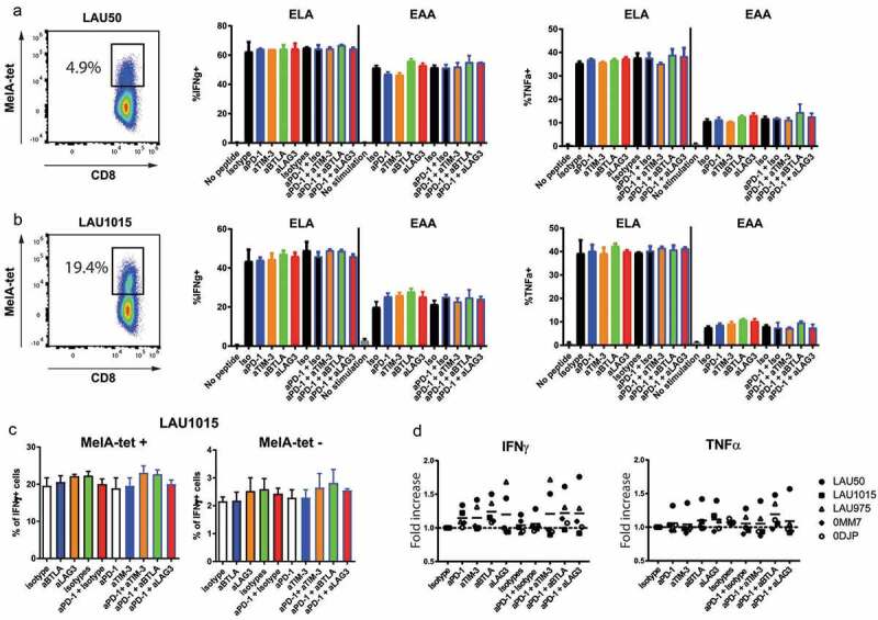
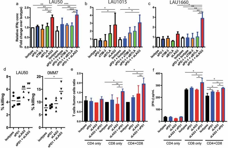
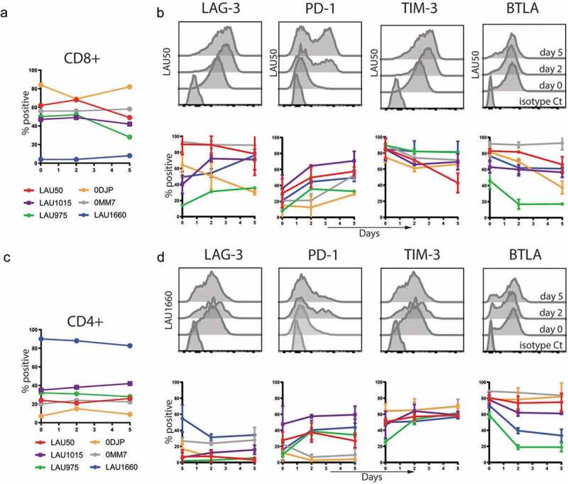
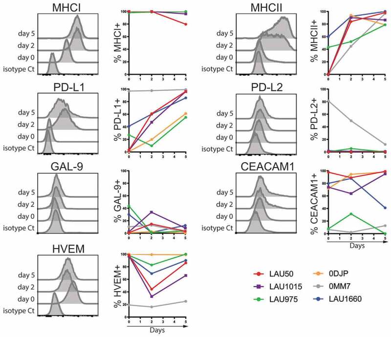
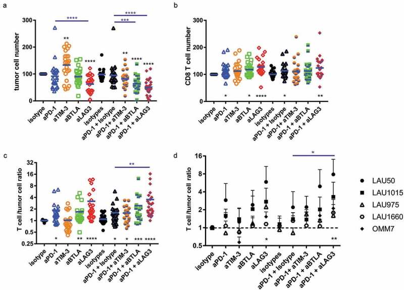
Despite the success of immunotherapy using checkpoint blockade, many patients with solid tumors remain refractory to these treatments. In human cancer, the experimental options to investigate the specific effects of antibodies blocking inhibitory receptors are limited and it is still unclear which cell types are involved. We addressed the question whether the direct interaction between T cells and tumor cells can be enforced through blocking a set of inhibitory receptors including PD-1, TIM-3, BTLA and LAG-3, blocked either individually or in dual combinations with the anti-PD-1 antibody, and to determine the condition that induces maximal T cell function preventing tumor cell proliferation. Using short-term Melan-A-specific or autologous re-stimulations, checkpoint blockade did not consistently increase cytokine production by tumor-derived expanded T cells. We next set up a 5-day co-culture assay with autologous melanoma cell lines and expanded tumor infiltrating T cells, originating from tumor specimens obtained from 6 different patients. Amongst all combos tested, we observed that blockade of LAG-3 alone, and more strongly when combined with PD-1 blockade, enforced T cell responses and tumor cell growth control. The combination of anti-LAG-3 plus anti-PD-1 acted through CD8 T cells and led to increased IFNγ production and cytotoxic capacity. Our results show that LAG-3 and PD-1 are regulating the direct interaction between tumor cells and autologous T cells, suggesting that therapy effects may be promoted by enhanced access of the corresponding blocking reagents to the tumor microenvironment.
尽管使用检查点阻断的免疫疗法取得了成功,但许多实体瘤患者对这些治疗仍有抗性。在人类癌症中,研究阻断抑制性受体的抗体的具体作用的实验选择有限,并且仍不清楚涉及哪些细胞类型。我们探讨了一个问题,即通过阻断一组抑制性受体(包括PD-1、TIM-3、BTLA和LAG-3),单独阻断或与抗PD-1抗体以双重组合的方式阻断,是否可以加强T细胞与肿瘤细胞之间的直接相互作用,并确定诱导最大T细胞功能以防止肿瘤细胞增殖的条件。使用短期的黑色素瘤抗原特异性或自体再刺激,检查点阻断并不能持续增加肿瘤来源的扩增T细胞产生的细胞因子。接下来,我们建立了一个为期5天的共培养试验,将自体黑色素瘤细胞系与扩增的肿瘤浸润T细胞共同培养,这些T细胞来自6名不同患者的肿瘤标本。在所有测试的组合中,我们观察到单独阻断LAG-3,以及与PD-1阻断联合时更强烈地阻断,可加强T细胞反应和肿瘤细胞生长控制。抗LAG-3加抗PD-1的组合通过CD8 T细胞起作用,并导致IFNγ产生增加和细胞毒性能力增强。我们的结果表明,LAG-3和PD-1正在调节肿瘤细胞与自体T细胞之间的直接相互作用,这表明相应阻断试剂更多地进入肿瘤微环境可能会促进治疗效果。