Javvaji Pradeep K, Dhali Arindam, Francis Joseph R, Kolte Atul P, Mech Anjumoni, Roy Sudhir C, Mishra Ashish, Bhatta Raghavendra
OMICS Laboratory, ICAR-National Institute of Animal Nutrition and Physiology, Bengaluru, India.
Center for Post Graduate Studies, Jain University, Bengaluru, India.
Front Cell Dev Biol. 2020 Aug 4;8:764. doi: 10.3389/fcell.2020.00764. eCollection 2020.
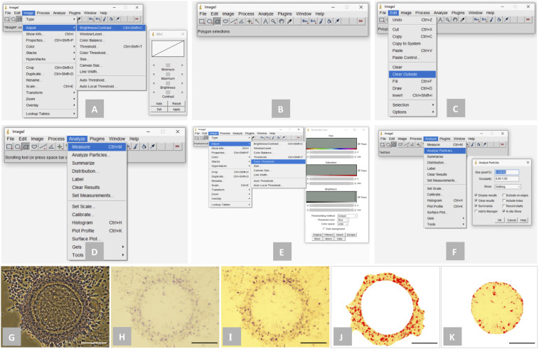
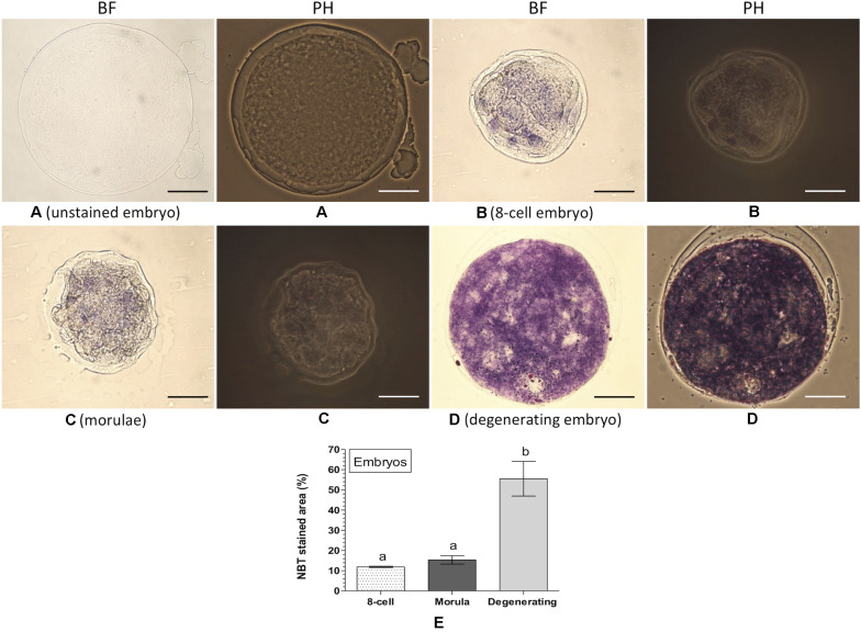
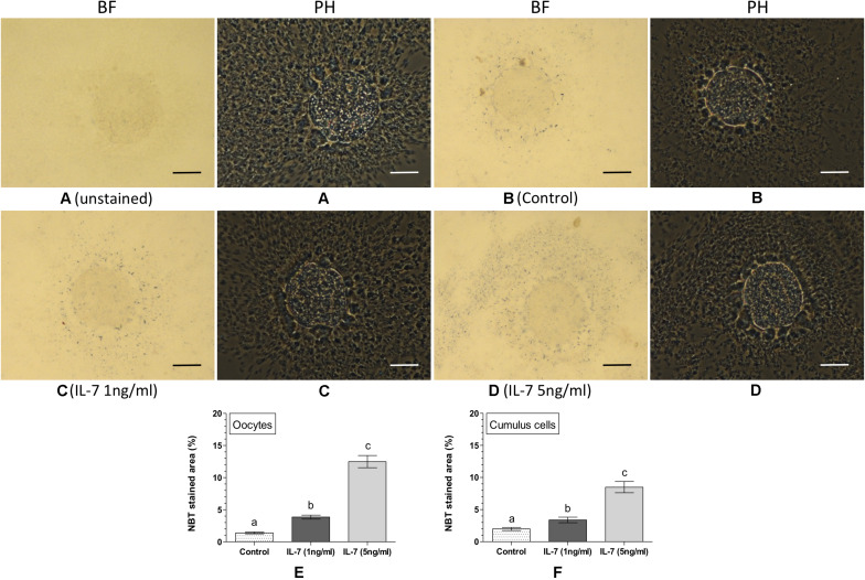
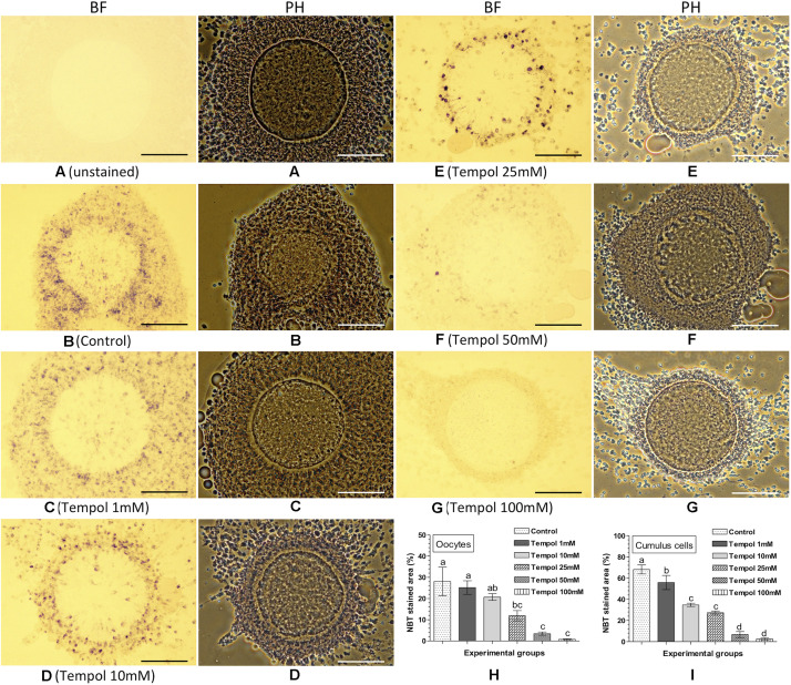
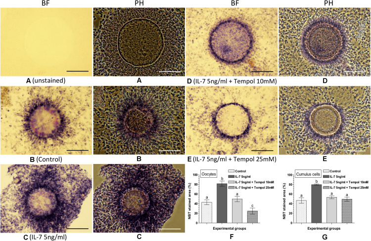
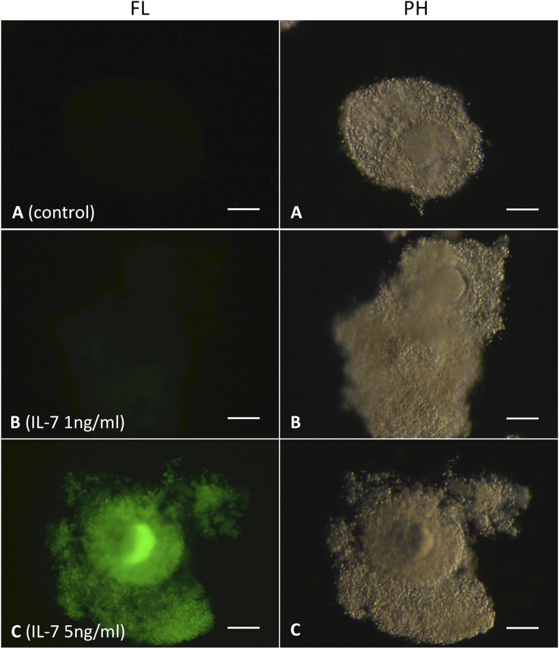
Assessment of intracellular reactive oxygen species (ROS) is important for evaluating the developmental ability of cumulus-oocyte complexes (COC) and embryos. Although, fluorescence-based 2',7'-dichlorodihydrofluorescein diacetate (DCFH-DA) staining method is used widely for detecting intracellular ROS in COC and embryos, it is associated with several limitations. This study aimed to develop an alternative method for detecting and quantifying intracellular ROS in oocytes, cumulus cells and embryos based on nitroblue tetrazolium (NBT) staining and bright-field microscopy. Nitroblue tetrazolium reacts with ROS and forms formazan precipitate that can be detected as dark purple/blue spots under bright-field microscope. Ovine COC were matured without (control) or with the supplementation of Interleukin-7 (IL-7; for stimulating intracellular ROS), Tempol (superoxide scavenger) or combination of IL-7 and Tempol. The matured COC were stained with NBT and the formation of intracellular formazan precipitates was assessed. Additionally, the matured COC were stained with DCFH-DA to compare the level of intracellular ROS. Further, ovine embryos (8-cell, morula, and degenerating) were generated and stained with NBT for assessing intracellular ROS. The level of intracellular ROS was expressed as the proportion (%) of the NBT stained area of oocytes, compact cumulus cell masses or embryos. The proportions of NBT stained area in the matured oocytes and cumulus cells was found significantly lesser in the control as compared to the IL-7 (1 and 5 ng/ml) treated groups. A similar trend in the intracellular ROS level was also observed in the matured COC, when assessed based on the DCFH-DA staining. Following the treatment with Tempol (100 mM), negligible NBT stained area in oocytes and cumulus cells was observed. The NBT staining patterns of the oocytes and cumulus cells following the combined treatment with IL-7 (5 ng/ml) and Tempol (10 and 25 mM) were comparable with that of the control. The proportion of NBT stained area did not differ significantly between the 8-cell embryos and morula, but was found significantly greater in the degenerating embryos. In conclusion, the developed NBT staining method was found effective for detecting and interpreting the level of intracellular ROS in oocytes, cumulus cells and embryos. This method can be used as an alternative to the DCFH-DA staining method.
评估细胞内活性氧(ROS)对于评估卵丘-卵母细胞复合体(COC)和胚胎的发育能力很重要。尽管基于荧光的2',7'-二氯二氢荧光素二乙酸酯(DCFH-DA)染色方法被广泛用于检测COC和胚胎中的细胞内ROS,但它存在一些局限性。本研究旨在开发一种基于硝基蓝四氮唑(NBT)染色和明场显微镜的方法,用于检测和定量卵母细胞、卵丘细胞和胚胎中的细胞内ROS。硝基蓝四氮唑与ROS反应形成甲臜沉淀,在明场显微镜下可检测为深紫色/蓝色斑点。绵羊COC在不添加(对照)或添加白细胞介素-7(IL-7;用于刺激细胞内ROS)、Tempol(超氧化物清除剂)或IL-7与Tempol组合的情况下成熟。成熟的COC用NBT染色,并评估细胞内甲臜沉淀的形成。此外,成熟的COC用DCFH-DA染色以比较细胞内ROS水平。进一步地,生成绵羊胚胎(8细胞、桑椹胚和退化胚胎)并用NBT染色以评估细胞内ROS。细胞内ROS水平以卵母细胞、致密卵丘细胞团或胚胎的NBT染色面积比例(%)表示。与IL-7(1和5 ng/ml)处理组相比,对照组中成熟卵母细胞和卵丘细胞的NBT染色面积比例显著较小。基于DCFH-DA染色评估时,成熟COC中的细胞内ROS水平也观察到类似趋势。用Tempol(100 mM)处理后,卵母细胞和卵丘细胞中观察到可忽略不计的NBT染色面积。IL-7(5 ng/ml)和Tempol(10和25 mM)联合处理后卵母细胞和卵丘细胞的NBT染色模式与对照组相当。8细胞胚胎和桑椹胚之间的NBT染色面积比例没有显著差异,但在退化胚胎中显著更大。总之,所开发的NBT染色方法被发现可有效检测和解释卵母细胞、卵丘细胞和胚胎中的细胞内ROS水平。该方法可作为DCFH-DA染色方法的替代方法。