Eleiwa Taher, Elsawy Amr, Özcan Eyüp, Abou Shousha Mohamed
Bascom Palmer Eye Institute, Miller School of Medicine, University of Miami, Miami, Florida 33136 USA.
Department of Ophthalmology, Faculty of Medicine, Benha University, Benha, Egypt.
Eye Vis (Lond). 2020 Sep 1;7:44. doi: 10.1186/s40662-020-00209-z. eCollection 2020.
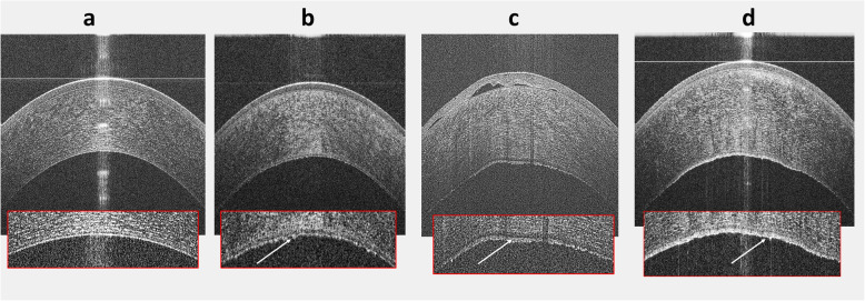
To describe the diagnostic performance of a deep learning algorithm in discriminating early-stage Fuchs' endothelial corneal dystrophy (FECD) without clinically evident corneal edema from healthy and late-stage FECD eyes using high-definition optical coherence tomography (HD-OCT).
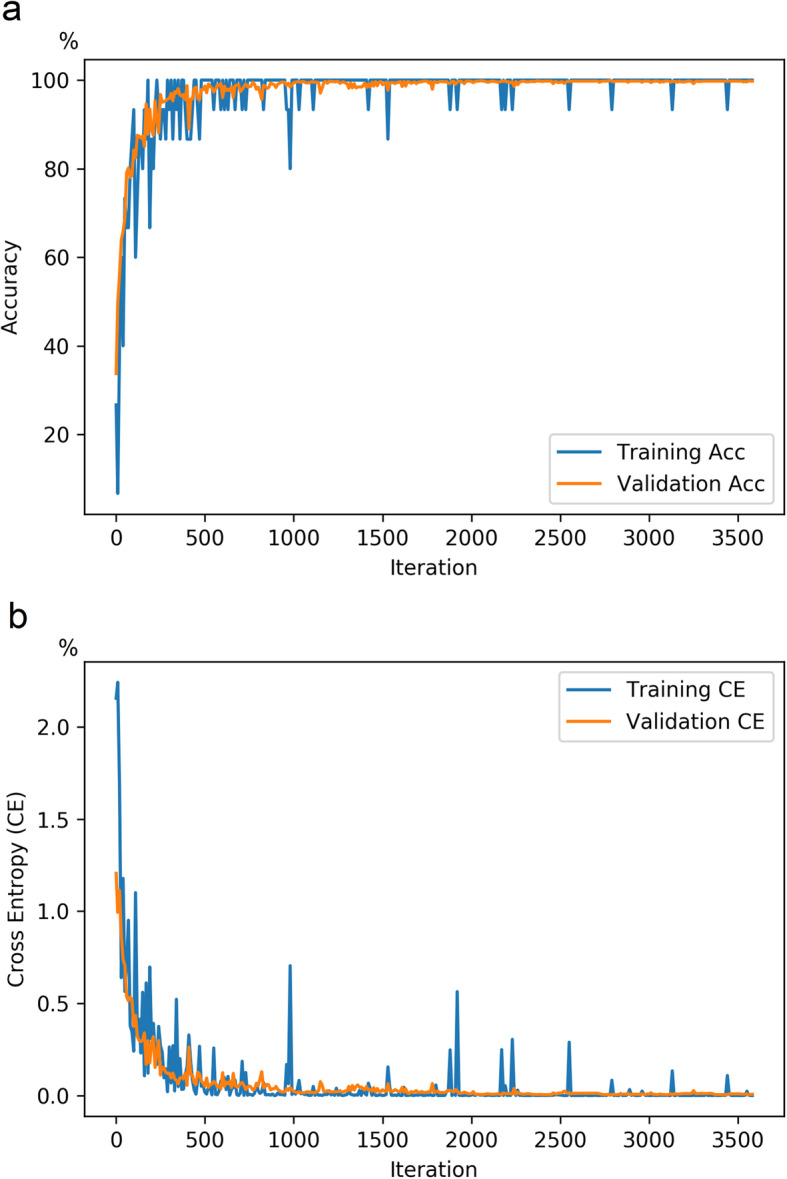
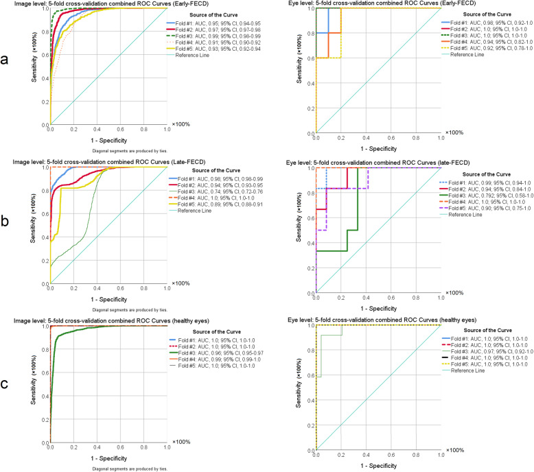
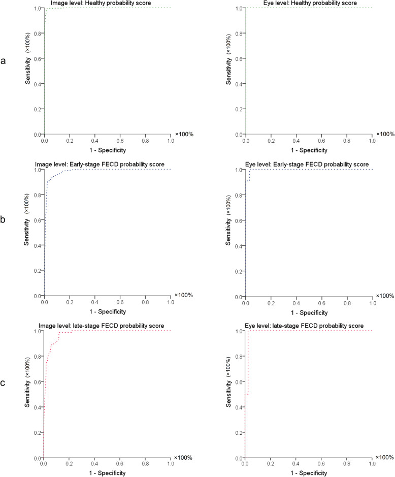
In this observational case-control study, 104 eyes (53 FECD eyes and 51 healthy controls) received HD-OCT imaging (Envisu R2210, Bioptigen, Buffalo Grove, IL, USA) using a 6 mm radial scan pattern centered on the corneal vertex. FECD was clinically categorized into early (without corneal edema) and late-stage (with corneal edema). A total of 18,720 anterior segment optical coherence tomography (AS-OCT) images (9180 healthy; 5400 early-stage FECD; 4140 late-stage FECD) of 104 eyes (81 patients) were used to develop and validate a deep learning classification network to differentiate early-stage FECD eyes from healthy eyes and those with clinical edema. Using 5-fold cross-validation on the dataset containing 11,340 OCT images (63 eyes), the network was trained with 80% of these images (3420 healthy; 3060 early-stage FECD; 2700 late-stage FECD), then tested with 20% (720 healthy; 720 early-stage FECD; 720 late-stage FECD). Thereafter, a final model was trained with the entire dataset consisting the 11,340 images and validated with a remaining 7380 images of unseen AS-OCT scans of 41 eyes (5040 healthy; 1620 early-stage FECD 720 late-stage FECD). Visualization of learned features was done, and area under curve (AUC), specificity, and sensitivity of the prediction outputs for healthy, early and late-stage FECD were computed.
The final model achieved an AUC of 0.997 ± 0.005 with 91% sensitivity and 97% specificity in detecting early-FECD; an AUC of 0.974 ± 0.005 with a specificity of 92% and a sensitivity up to 100% in detecting late-stage FECD; and an AUC of 0.998 ± 0.001 with a specificity 98% and a sensitivity of 99% in discriminating healthy corneas from all FECD.
Deep learning algorithm is an accurate autonomous novel diagnostic tool of FECD with very high sensitivity and specificity that can be used to grade FECD severity with high accuracy.
使用高清光学相干断层扫描(HD-OCT)描述深度学习算法在区分无临床明显角膜水肿的早期富克斯内皮性角膜营养不良(FECD)与健康及晚期FECD眼方面的诊断性能。
在这项观察性病例对照研究中,104只眼(53只FECD眼和51只健康对照)接受了HD-OCT成像(Envisu R2210,Bioptigen,美国伊利诺伊州布法罗格罗夫),采用以角膜顶点为中心的6毫米径向扫描模式。FECD在临床上分为早期(无角膜水肿)和晚期(有角膜水肿)。对104只眼(81例患者)的总共18720张眼前节光学相干断层扫描(AS-OCT)图像(9180张健康图像;5400张早期FECD图像;4140张晚期FECD图像)进行分析,以开发和验证一个深度学习分类网络,用于区分早期FECD眼与健康眼以及有临床水肿的眼。在包含11340张OCT图像(63只眼)的数据集上进行5折交叉验证,该网络用这些图像的80%(3420张健康图像;3060张早期FECD图像;2700张晚期FECD图像)进行训练,然后用20%(720张健康图像;720张早期FECD图像;720张晚期FECD图像)进行测试。此后,使用由这11340张图像组成的整个数据集训练最终模型,并用另外41只眼(5040张健康图像;1620张早期FECD图像;720张晚期FECD图像)的7380张未见过的AS-OCT扫描图像进行验证。对学习到的特征进行可视化,并计算健康、早期和晚期FECD预测输出的曲线下面积(AUC)、特异性和敏感性。
最终模型在检测早期FECD时AUC为0.997±0.005,敏感性为91%,特异性为97%;在检测晚期FECD时AUC为0.974±0.005,特异性为92%,敏感性高达100%;在区分健康角膜与所有FECD时AUC为0.998±0.001,特异性为98%,敏感性为99%。
深度学习算法是一种准确的自主新型FECD诊断工具,具有非常高的敏感性和特异性,可用于高精度分级FECD严重程度。