• 文献检索
  • 文档翻译
  • 深度研究
  • 学术资讯
  • Suppr Zotero 插件Zotero 插件
  • 邀请有礼
  • 套餐&价格
  • 历史记录
应用&插件
Suppr Zotero 插件Zotero 插件浏览器插件Mac 客户端Windows 客户端微信小程序
定价
高级版会员购买积分包购买API积分包
服务
文献检索文档翻译深度研究API 文档MCP 服务
关于我们
关于 Suppr公司介绍联系我们用户协议隐私条款
关注我们

Suppr 超能文献

核心技术专利:CN118964589B侵权必究
粤ICP备2023148730 号-1Suppr @ 2026

文献检索

告别复杂PubMed语法,用中文像聊天一样搜索,搜遍4000万医学文献。AI智能推荐,让科研检索更轻松。

立即免费搜索

文件翻译

保留排版,准确专业,支持PDF/Word/PPT等文件格式,支持 12+语言互译。

免费翻译文档

深度研究

AI帮你快速写综述,25分钟生成高质量综述,智能提取关键信息,辅助科研写作。

立即免费体验

60 例新型冠状病毒病(COVID-19)患者的胸部计算机断层扫描(CT)表现和半定量评分:解剖与病理相结合的回顾性影像学分析。

Chest computed tomography (CT) findings and semiquantitative scoring of 60 patients with coronavirus disease 2019 (COVID-19): A retrospective imaging analysis combining anatomy and pathology.

机构信息

Department of Radiology, Dianjiang People's Hospital of Chongqing, Chongqing, China.

Department of General Surgery, Dianjiang People's Hospital of Chongqing, Chongqing, China.

出版信息

PLoS One. 2020 Sep 4;15(9):e0238760. doi: 10.1371/journal.pone.0238760. eCollection 2020.

DOI:10.1371/journal.pone.0238760
PMID:32886711
原文链接:https://pmc.ncbi.nlm.nih.gov/articles/PMC7473568/
Abstract

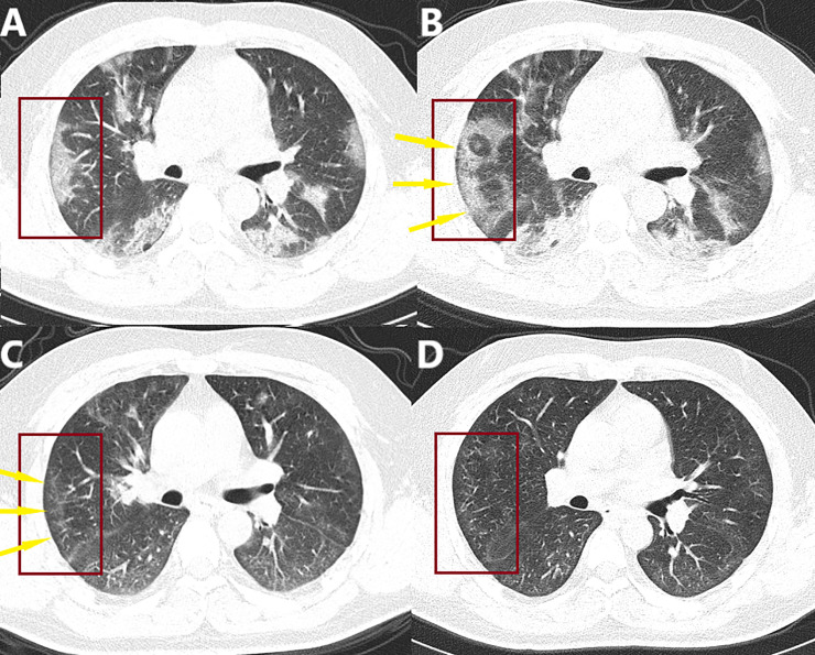
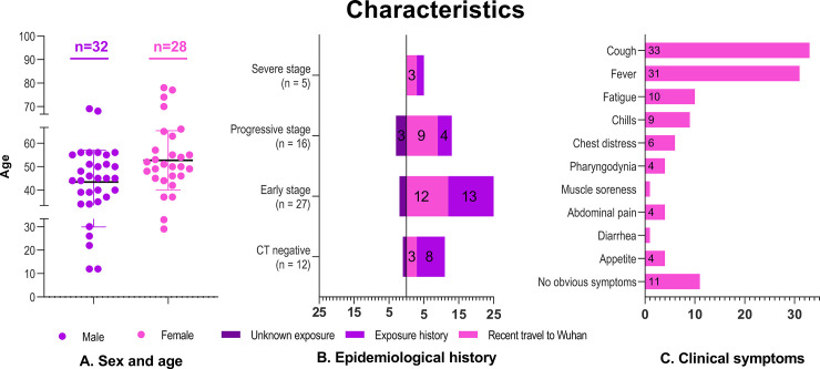
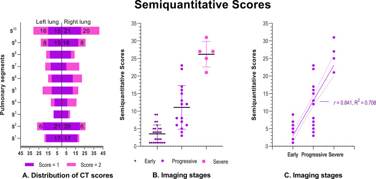
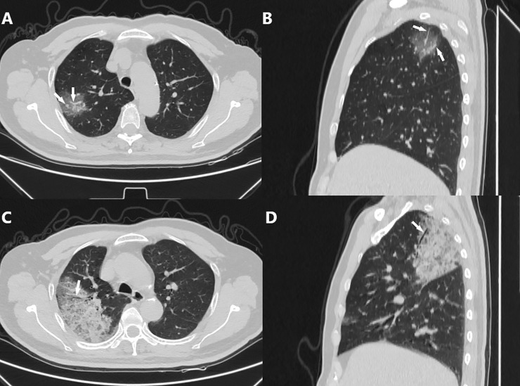
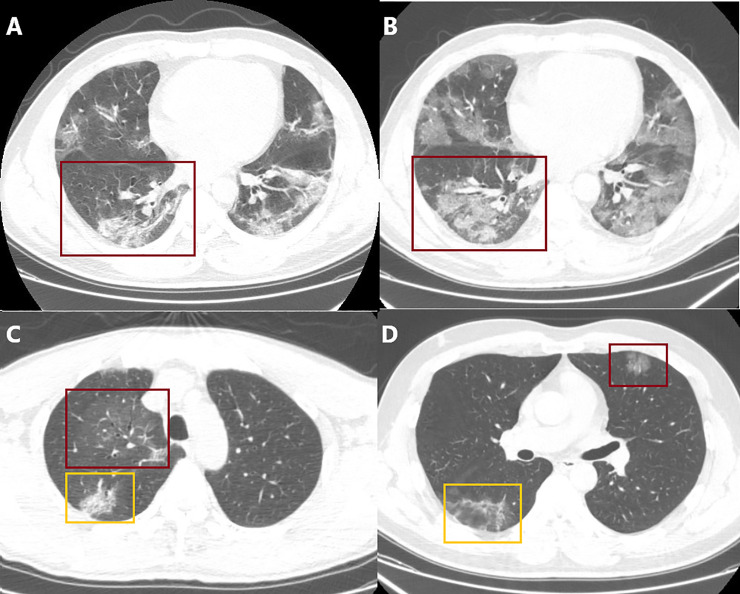
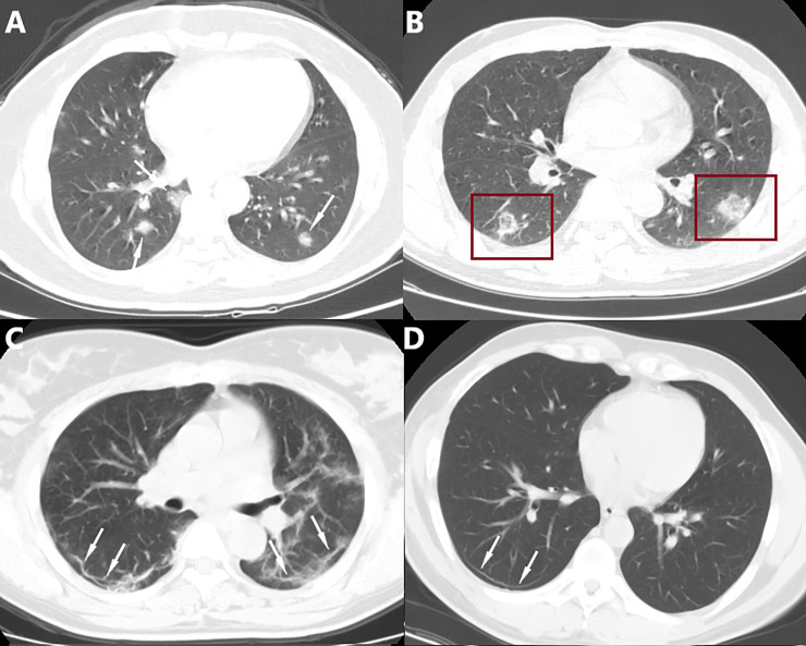
In this study, we ascertained the chest CT data of 60 patients admitted to 3 hospitals in Chongqing with confirmed COVID-19. We conducted anatomical and pathological analyses to elucidate the possible reasons for the distribution, morphology, and characteristics of COVID-19 in chest CT. We also shared a semiquantitative scoring of affected lung segments, which was recommended by our local medical association. This scoring system was applied to quantify the severity of the disease. The most frequent imaging findings of COVID-19 were subpleural ground glass opacities and consolidation; there was a significant difference in semiquantitative scores between the early, progressive, and severe stages of the disease. We conclude that the chest CT findings of COVID-19 showed certain characteristics because of the anatomical features of the human body and pathological changes caused by the virus. Therefore, chest CT is a valuable tool for facilitating the diagnosis of COVID-19 and semiquantitative scoring of affected lung segments may further elucidate diagnosis and assessment of disease severity. This will assist healthcare workers in diagnosing COVID-19 and assessing disease severity, facilitate the selection of appropriate treatment options, which is important for reducing the spread of the virus, saving lives, and controlling the pandemic.

摘要

在这项研究中,我们确定了来自重庆 3 家医院的 60 名确诊 COVID-19 患者的胸部 CT 数据。我们进行了解剖学和病理学分析,以阐明 COVID-19 在胸部 CT 中的分布、形态和特征的可能原因。我们还分享了当地医学会推荐的受累肺段的半定量评分,该评分系统用于量化疾病的严重程度。COVID-19 的最常见影像学表现是胸膜下磨玻璃影和实变;疾病的早期、进展期和严重期之间的半定量评分有显著差异。我们的结论是,由于人体的解剖学特征和病毒引起的病理学变化,COVID-19 的胸部 CT 表现具有一定的特征。因此,胸部 CT 是有助于 COVID-19 诊断的有价值的工具,受累肺段的半定量评分可能进一步阐明诊断和疾病严重程度评估。这将有助于医疗保健工作者诊断 COVID-19 和评估疾病严重程度,促进选择合适的治疗方案,这对于减少病毒传播、拯救生命和控制大流行非常重要。

https://cdn.ncbi.nlm.nih.gov/pmc/blobs/685a/7473568/6ec95de579db/pone.0238760.g008.jpg
https://cdn.ncbi.nlm.nih.gov/pmc/blobs/685a/7473568/09763a6a6d54/pone.0238760.g001.jpg
https://cdn.ncbi.nlm.nih.gov/pmc/blobs/685a/7473568/93892f724f2f/pone.0238760.g002.jpg
https://cdn.ncbi.nlm.nih.gov/pmc/blobs/685a/7473568/5441ef2ac7c6/pone.0238760.g003.jpg
https://cdn.ncbi.nlm.nih.gov/pmc/blobs/685a/7473568/bd4c5045b30b/pone.0238760.g004.jpg
https://cdn.ncbi.nlm.nih.gov/pmc/blobs/685a/7473568/afb4bfca00ab/pone.0238760.g005.jpg
https://cdn.ncbi.nlm.nih.gov/pmc/blobs/685a/7473568/c825e5071b14/pone.0238760.g006.jpg
https://cdn.ncbi.nlm.nih.gov/pmc/blobs/685a/7473568/2613473937a0/pone.0238760.g007.jpg
https://cdn.ncbi.nlm.nih.gov/pmc/blobs/685a/7473568/6ec95de579db/pone.0238760.g008.jpg

相似文献

1
Chest computed tomography (CT) findings and semiquantitative scoring of 60 patients with coronavirus disease 2019 (COVID-19): A retrospective imaging analysis combining anatomy and pathology.60 例新型冠状病毒病(COVID-19)患者的胸部计算机断层扫描(CT)表现和半定量评分:解剖与病理相结合的回顾性影像学分析。
PLoS One. 2020 Sep 4;15(9):e0238760. doi: 10.1371/journal.pone.0238760. eCollection 2020.
2
The role of a chest computed tomography severity score in coronavirus disease 2019 pneumonia.胸部计算机断层扫描严重程度评分在2019冠状病毒病肺炎中的作用
Medicine (Baltimore). 2020 Oct 16;99(42):e22433. doi: 10.1097/MD.0000000000022433.
3
CT image of novel coronavirus pneumonia: a case report.新型冠状病毒肺炎的CT图像:病例报告
Jpn J Radiol. 2020 May;38(5):407-408. doi: 10.1007/s11604-020-00945-1. Epub 2020 Mar 18.
4
A retrospective study of the initial chest CT imaging findings in 50 COVID-19 patients stratified by gender and age.50 例 COVID-19 患者按性别和年龄分层的初始胸部 CT 影像学表现的回顾性研究。
J Xray Sci Technol. 2020;28(5):875-884. doi: 10.3233/XST-200709.
5
Time Course of Lung Changes at Chest CT during Recovery from Coronavirus Disease 2019 (COVID-19).新冠肺炎(COVID-19)康复过程中胸部 CT 肺部变化的时间进程。
Radiology. 2020 Jun;295(3):715-721. doi: 10.1148/radiol.2020200370. Epub 2020 Feb 13.
6
Preliminary CT findings of coronavirus disease 2019 (COVID-19).新型冠状病毒肺炎(COVID-19)的初步 CT 影像学表现。
Clin Imaging. 2020 Sep;65:124-132. doi: 10.1016/j.clinimag.2020.04.042. Epub 2020 May 12.
7
CT in coronavirus disease 2019 (COVID-19): a systematic review of chest CT findings in 4410 adult patients.CT 在 2019 年冠状病毒病(COVID-19)中的应用:对 4410 例成年患者胸部 CT 表现的系统综述。
Eur Radiol. 2020 Nov;30(11):6129-6138. doi: 10.1007/s00330-020-06975-7. Epub 2020 May 30.
8
Chest CT Findings in Patients With Coronavirus Disease 2019 and Its Relationship With Clinical Features.新型冠状病毒肺炎患者的胸部 CT 表现及其与临床特征的关系。
Invest Radiol. 2020 May;55(5):257-261. doi: 10.1097/RLI.0000000000000670.
9
Chest CT features and progression of patients with coronavirus disease 2019.2019冠状病毒病患者的胸部CT特征及病情进展
Br J Radiol. 2020 Dec 1;93(1116):20200219. doi: 10.1259/bjr.20200219. Epub 2020 Nov 13.
10
The Performance of Chest CT in Evaluating the Clinical Severity of COVID-19 Pneumonia: Identifying Critical Cases Based on CT Characteristics.胸部 CT 对评估 COVID-19 肺炎临床严重程度的性能:基于 CT 特征识别重症病例。
Invest Radiol. 2020 Jul;55(7):412-421. doi: 10.1097/RLI.0000000000000689.

引用本文的文献

1
Predicting malignant potential of solitary pulmonary nodules in patients with COVID-19 infection: a comprehensive analysis of CT imaging and tumor markers.预测 COVID-19 感染患者孤立性肺结节的恶性潜能:CT 影像学与肿瘤标志物的综合分析。
BMC Infect Dis. 2024 Sep 27;24(1):1050. doi: 10.1186/s12879-024-09952-3.
2
Association of chest computed tomography severity score at ICU admission and respiratory outcomes in critically ill COVID-19 patients.入住 ICU 时的胸部 CT 严重程度评分与危重症 COVID-19 患者的呼吸结局的相关性。
PLoS One. 2024 May 2;19(5):e0299390. doi: 10.1371/journal.pone.0299390. eCollection 2024.

本文引用的文献

1
Chest CT score in COVID-19 patients: correlation with disease severity and short-term prognosis.COVID-19 患者的胸部 CT 评分:与疾病严重程度和短期预后的相关性。
Eur Radiol. 2020 Dec;30(12):6808-6817. doi: 10.1007/s00330-020-07033-y. Epub 2020 Jul 4.
2
[Analysis of coronavirus disease-19 (COVID-19) based on SARS autopsy].基于严重急性呼吸综合征(SARS)尸检的新型冠状病毒肺炎(COVID-19)分析
Zhonghua Bing Li Xue Za Zhi. 2020 Apr 8;49(4):291-293. doi: 10.3760/cma.j.cn112151-20200211-00114.
3
CT image visual quantitative evaluation and clinical classification of coronavirus disease (COVID-19).
CT 影像视觉定量评估及新型冠状病毒肺炎(COVID-19)临床分类。
Eur Radiol. 2020 Aug;30(8):4407-4416. doi: 10.1007/s00330-020-06817-6. Epub 2020 Mar 25.
4
Coronavirus disease 2019: initial chest CT findings.新型冠状病毒肺炎的胸部 CT 表现。
Eur Radiol. 2020 Aug;30(8):4398-4406. doi: 10.1007/s00330-020-06816-7. Epub 2020 Mar 24.
5
Gross examination report of a COVID-19 death autopsy.一份新冠死亡病例尸检大体检查报告。
Fa Yi Xue Za Zhi. 2020 Feb;36(1):21-23. doi: 10.12116/j.issn.1004-5619.2020.01.005. Epub 2020 Feb 25.
6
Review and Prospect of Pathological Features of Corona Virus Disease.冠状病毒病病理特征的回顾与展望
Fa Yi Xue Za Zhi. 2020 Feb;36(1):16-20. doi: 10.12116/j.issn.1004-5619.2020.01.004. Epub 2020 Feb 25.
7
Chest CT manifestations of new coronavirus disease 2019 (COVID-19): a pictorial review.新型冠状病毒病 2019(COVID-19)的胸部 CT 表现:影像学综述。
Eur Radiol. 2020 Aug;30(8):4381-4389. doi: 10.1007/s00330-020-06801-0. Epub 2020 Mar 19.
8
Temporal Changes of CT Findings in 90 Patients with COVID-19 Pneumonia: A Longitudinal Study.90 例 COVID-19 肺炎患者 CT 表现的时间变化:一项纵向研究。
Radiology. 2020 Aug;296(2):E55-E64. doi: 10.1148/radiol.2020200843. Epub 2020 Mar 19.
9
[A pathological report of three COVID-19 cases by minimal invasive autopsies].[三例新冠病毒病病例微创尸检病理报告]
Zhonghua Bing Li Xue Za Zhi. 2020 May 8;49(5):411-417. doi: 10.3760/cma.j.cn112151-20200312-00193.
10
CT Features of Coronavirus Disease 2019 (COVID-19) Pneumonia in 62 Patients in Wuhan, China.中国武汉 62 例 2019 年冠状病毒病(COVID-19)肺炎的 CT 特征。
AJR Am J Roentgenol. 2020 Jun;214(6):1287-1294. doi: 10.2214/AJR.20.22975. Epub 2020 Mar 5.