Department of Radiology, the Second Affiliated Hospital of Chongqing Medical University, Chongqing, China.
Department of Radiology, Chongqing Three Gorges Central Hospital, Chongqing, 404000, China.
Eur Radiol. 2020 Aug;30(8):4398-4406. doi: 10.1007/s00330-020-06816-7. Epub 2020 Mar 24.
To systematically analyze CT findings during the early and progressive stages of natural course of coronavirus disease 2019 and also to explore possible changes in pulmonary parenchymal abnormalities during these two stages.
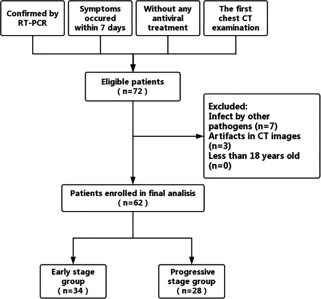
We retrospectively reviewed the initial chest CT data of 62 confirmed coronavirus disease 2019 patients (34 men, 28 women; age range 20-91 years old) who did not receive any antiviral treatment between January 21 and February 4, 2020, in Chongqing, China. Patients were assigned to the early-stage group (onset of symptoms within 4 days) or progressive-stage group (onset of symptoms within 4-7 days) for analysis. CT characteristics and the distribution, size, and CT score of pulmonary parenchymal abnormalities were assessed.
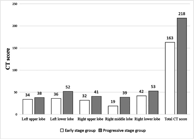
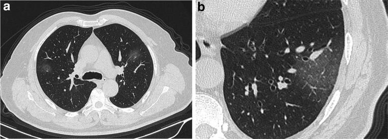
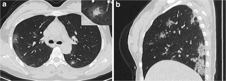
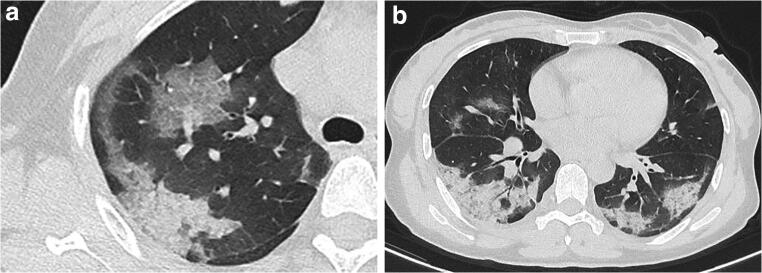
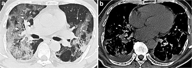
In our study, the major characteristic of coronavirus disease 2019 was ground-glass opacity (61.3%), followed by ground-glass opacity with consolidation (35.5%), rounded opacities (25.8%), a crazy-paving pattern (25.8%), and an air bronchogram (22.6%). No patient presented cavitation, a reticular pattern, or bronchial wall thickening. The CT scores of the progressive-stage group were significantly greater than those of the early-stage group (p = 0.004).
Multiple ground-glass opacities with consolidations in the periphery of the lungs were the primary CT characteristic of coronavirus disease 2019. CT score can be used to evaluate the severity of the disease. If these typical alterations are found, then the differential diagnosis of coronavirus disease 2019 must be considered.
• Multiple GGOs with consolidations in the periphery of the lungs were the primary CT characteristic of COVID-19. • The halo sign may be a special CT feature in the early-stage COVID-19 patients. • Significantly increased CT score may indicate the aggravation of COVID-19 in the progressive stage.
系统分析 2019 年冠状病毒病自然病程的早期和进展阶段的 CT 表现,并探讨这两个阶段肺实质异常的可能变化。
我们回顾性分析了 2020 年 1 月 21 日至 2 月 4 日期间在中国重庆未接受任何抗病毒治疗的 62 例确诊的 2019 年冠状病毒病患者的初始胸部 CT 数据(34 名男性,28 名女性;年龄 20-91 岁)。患者被分为早期组(症状发作 4 天内)或进展期组(症状发作 4-7 天内)进行分析。评估 CT 特征以及肺实质异常的分布、大小和 CT 评分。
在我们的研究中,2019 年冠状病毒病的主要特征是磨玻璃影(61.3%),其次是磨玻璃影伴实变(35.5%)、类圆形阴影(25.8%)、铺路石征(25.8%)和空气支气管征(22.6%)。没有患者出现空洞、网状模式或支气管壁增厚。进展期组的 CT 评分明显高于早期组(p=0.004)。
肺部周边多发磨玻璃影伴实变是 2019 年冠状病毒病的主要 CT 特征。CT 评分可用于评估疾病的严重程度。如果发现这些典型改变,则必须考虑 2019 年冠状病毒病的鉴别诊断。
肺部周边多发磨玻璃影伴实变是 COVID-19 的主要 CT 特征。
晕征可能是 COVID-19 早期的一个特殊 CT 特征。
进展期 COVID-19 患者 CT 评分显著增加可能提示病情加重。