Queen Square Multiple Sclerosis Centre, Department of Neuroinflammation, UCL Queen Square Institute of Neurology, University College London, London, UK/Unità di neurologia, Associazione Centro 'Dino Ferrari', IRCCS Fondazione Ca' Granda Ospedale Maggiore Policlinico, University of Milan, Milan, Italy.
Queen Square Multiple Sclerosis Centre, Department of Neuroinflammation, UCL Queen Square Institute of Neurology, University College London, London, UK/Department of Biomedical Imaging and Image-Guided Therapy, Medical University of Vienna, Vienna, Austria.
Mult Scler. 2022 Apr;28(5):683-690. doi: 10.1177/1352458520958589. Epub 2020 Sep 23.
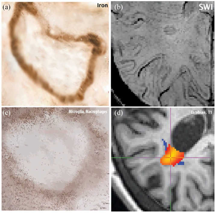
New clinical activity in multiple sclerosis (MS) is often accompanied by acute inflammation which subsides. However, there is growing evidence that a substantial proportion of lesions remain active well beyond the acute phase. Chronic active lesions are most frequently found in progressive MS and are characterised by a border of inflammation associated with iron-enriched cells, leading to ongoing tissue injury. Identifying imaging markers for chronic active lesions in vivo are thus a major research goal. We reviewed the literature on imaging of chronic active lesion in MS, focussing on 'slowly expanding lesions' (SELs), detected by volumetric longitudinal magnetic resonance imaging (MRI) and 'rim-positive' lesions, identified by susceptibility iron-sensitive MRI. Both SELs and rim-positive lesions have been found to be prognostically relevant to future disability. Little is known about the co-occurrence of rims around SELs and their inter-relationship with other emerging techniques such as dynamic contrast enhancement (DCE) and positron emission tomography (PET).
多发性硬化症(MS)中的新临床活动通常伴随着消退的急性炎症。然而,越来越多的证据表明,很大一部分病变在急性期过后仍然活跃。慢性活动性病变最常见于进展性 MS 中,其特征是炎症边界与富含铁的细胞相关,导致持续的组织损伤。因此,确定体内慢性活动性病变的成像标志物是一个主要的研究目标。我们回顾了多发性硬化症慢性活动性病变的影像学研究文献,重点关注体积纵向磁共振成像(MRI)检测到的“缓慢扩张病变”(SELs)和磁化率敏感 MRI 检测到的“边缘阳性病变”。SELs 和边缘阳性病变均与未来残疾的预后相关。关于 SELs 周围边缘的共存及其与其他新兴技术(如动态对比增强(DCE)和正电子发射断层扫描(PET))之间的相互关系,人们知之甚少。