Department of Cell Biology, Johns Hopkins University, School of Medicine, Baltimore, MD, USA.
Biochemistry, Cellular and Molecular Biology Graduate Program, Johns Hopkins University, School of Medicine, Baltimore, MD, USA.
Nat Neurosci. 2020 Nov;23(11):1329-1338. doi: 10.1038/s41593-020-00716-1. Epub 2020 Sep 28.
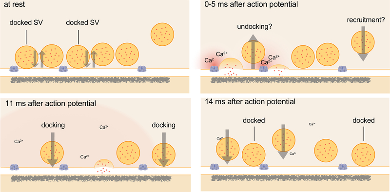
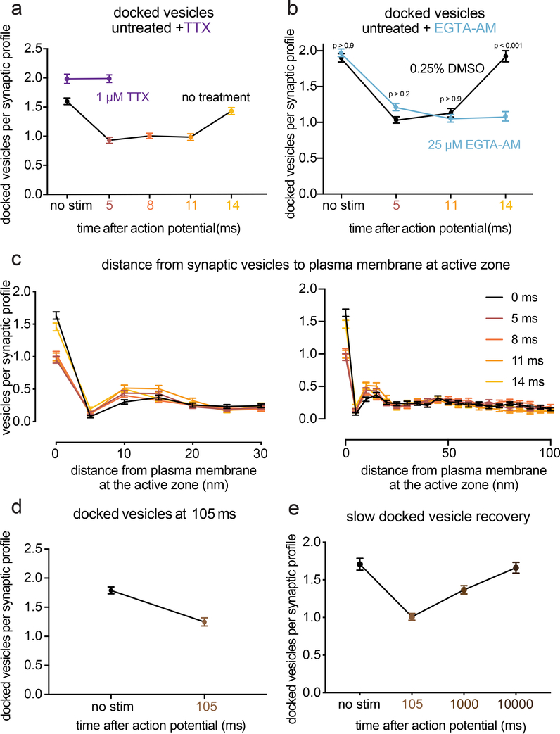
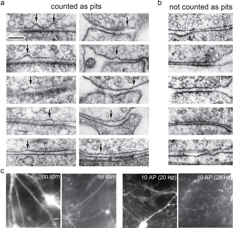
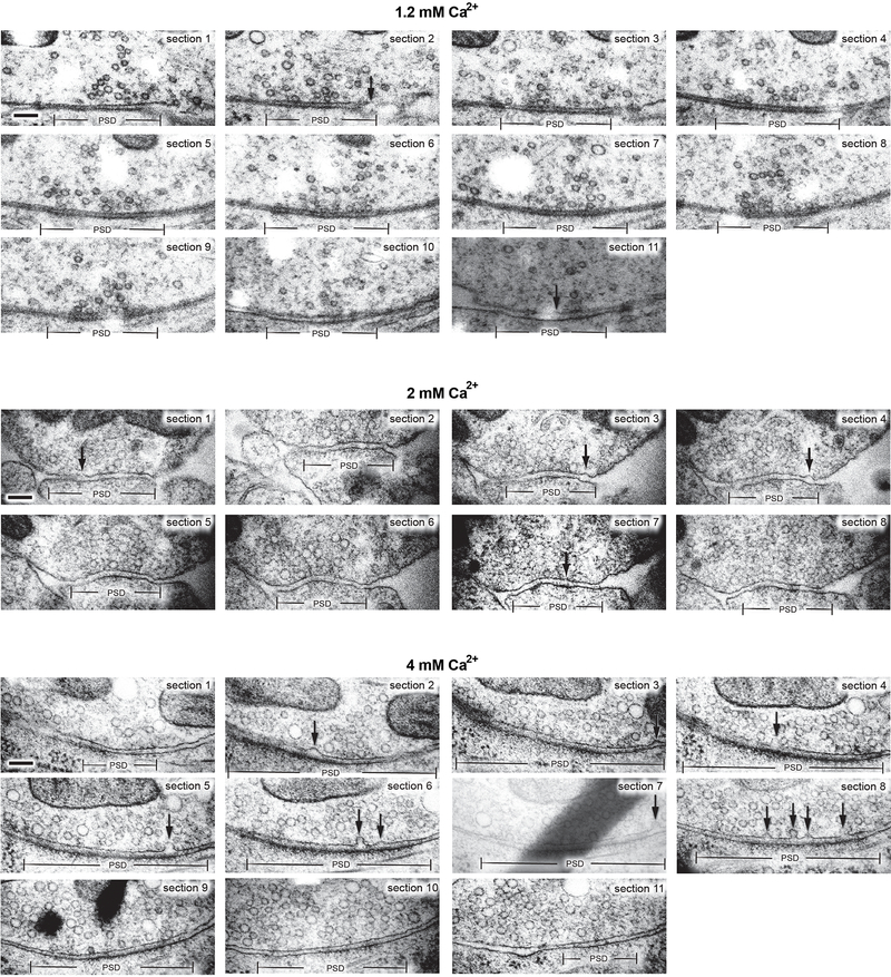
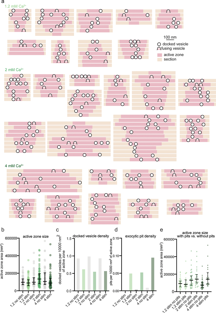
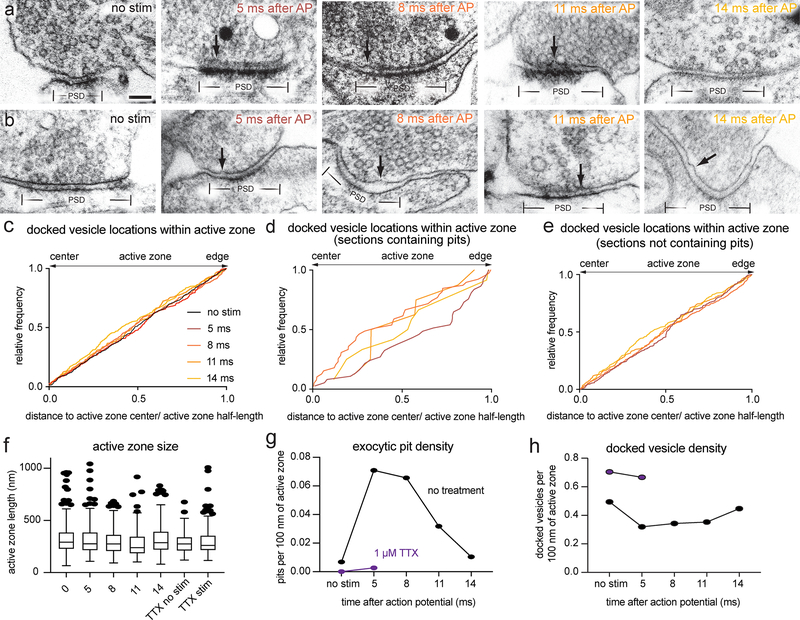
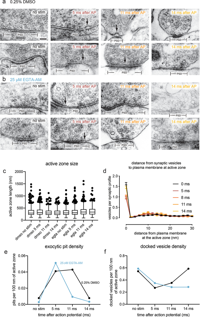
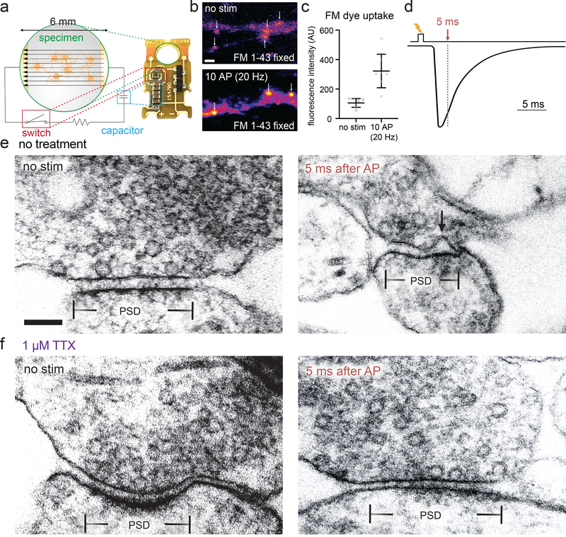
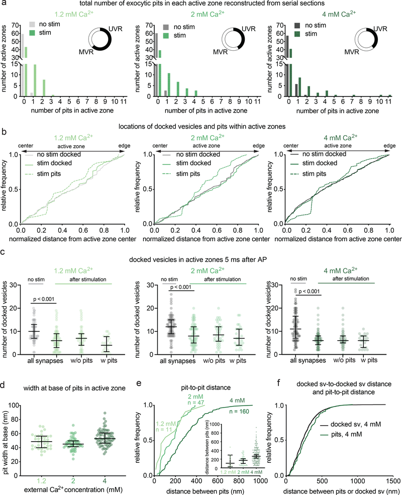
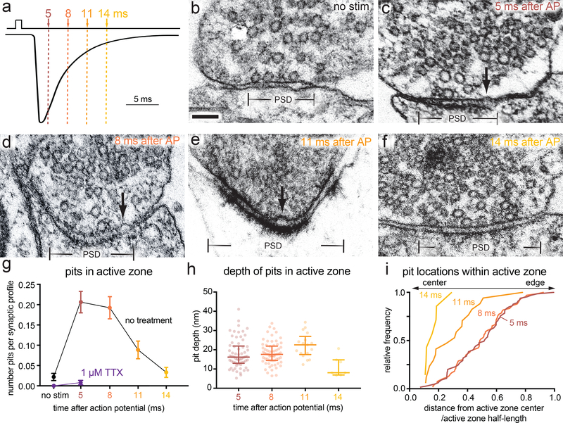
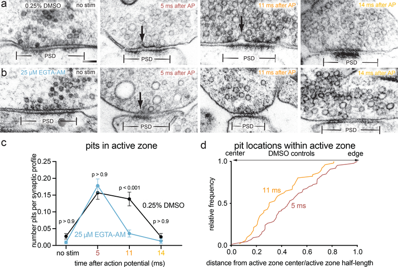
Synaptic vesicles fuse with the plasma membrane to release neurotransmitter following an action potential, after which new vesicles must 'dock' to refill vacated release sites. To capture synaptic vesicle exocytosis at cultured mouse hippocampal synapses, we induced single action potentials by electrical field stimulation, then subjected neurons to high-pressure freezing to examine their morphology by electron microscopy. During synchronous release, multiple vesicles can fuse at a single active zone. Fusions during synchronous release are distributed throughout the active zone, whereas fusions during asynchronous release are biased toward the center of the active zone. After stimulation, the total number of docked vesicles across all synapses decreases by ~40%. Within 14 ms, new vesicles are recruited and fully replenish the docked pool, but this docking is transient and they either undock or fuse within 100 ms. These results demonstrate that the recruitment of synaptic vesicles to release sites is rapid and reversible.
动作电位引发突触囊泡融合并释放神经递质,之后新的囊泡必须“停靠”到空出的释放位点来进行补充。为了在体外培养的海马神经元突触上捕获突触囊泡胞吐的过程,我们通过电场刺激诱发单个动作电位,然后对神经元进行高压冷冻,通过电子显微镜检查它们的形态。在同步释放期间,多个囊泡可以在单个活性区融合。在同步释放过程中,融合发生在活性区的各个部位,而在异步释放过程中,融合更偏向活性区的中心。刺激后,所有突触的停靠囊泡总数减少了约 40%。在 14 毫秒内,新的囊泡被募集并完全补充停靠池,但这种停靠是短暂的,它们在 100 毫秒内要么脱附,要么融合。这些结果表明,囊泡向释放位点的募集是快速和可逆的。