• 文献检索
  • 文档翻译
  • 深度研究
  • 学术资讯
  • Suppr Zotero 插件Zotero 插件
  • 邀请有礼
  • 套餐&价格
  • 历史记录
应用&插件
Suppr Zotero 插件Zotero 插件浏览器插件Mac 客户端Windows 客户端微信小程序
定价
高级版会员购买积分包购买API积分包
服务
文献检索文档翻译深度研究API 文档MCP 服务
关于我们
关于 Suppr公司介绍联系我们用户协议隐私条款
关注我们

Suppr 超能文献

核心技术专利:CN118964589B侵权必究
粤ICP备2023148730 号-1Suppr @ 2026

文献检索

告别复杂PubMed语法,用中文像聊天一样搜索,搜遍4000万医学文献。AI智能推荐,让科研检索更轻松。

立即免费搜索

文件翻译

保留排版,准确专业,支持PDF/Word/PPT等文件格式,支持 12+语言互译。

免费翻译文档

深度研究

AI帮你快速写综述,25分钟生成高质量综述,智能提取关键信息,辅助科研写作。

立即免费体验

相关光镜和透射电镜显示了丙酸处理的SH-SY5Y细胞中线粒体质量控制介导的线粒体自噬细节。

Correlative Light and Transmission Electron Microscopy Showed Details of Mitophagy by Mitochondria Quality Control in Propionic Acid Treated SH-SY5Y Cell.

作者信息

Jung Minkyo, Choi Hyosun, Kim Jaekwang, Mun Ji Young

机构信息

Neural Circuit Research Group, Korea Brain Research Institute, Daegu 41062, Korea.

BK21 Plus Program, Department of Senior Healthcare, Graduate School, Eulji University, Daejeon 34824, Korea.

出版信息

Materials (Basel). 2020 Sep 29;13(19):4336. doi: 10.3390/ma13194336.

DOI:10.3390/ma13194336
PMID:33003589
原文链接:https://pmc.ncbi.nlm.nih.gov/articles/PMC7579125/
Abstract

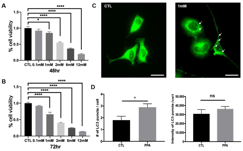
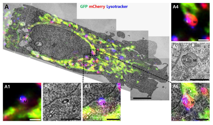
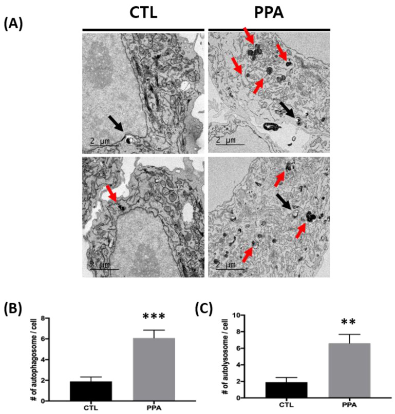
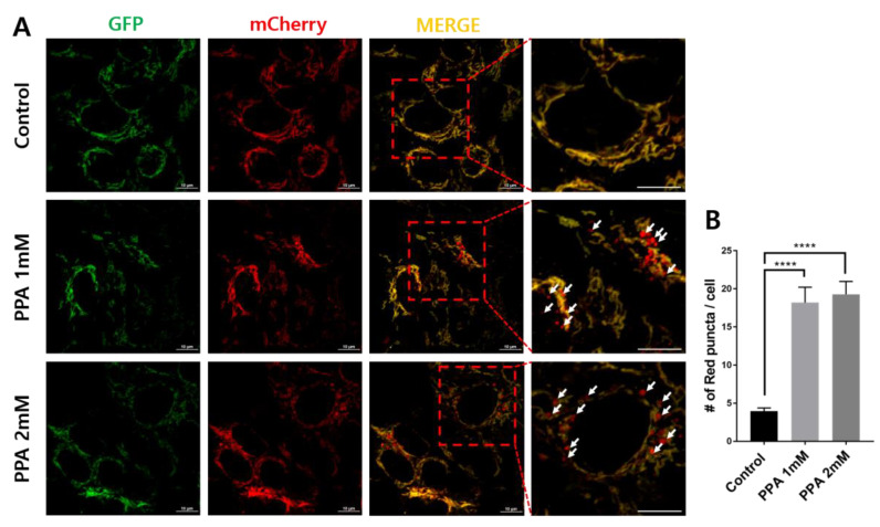
Propionic acid is a metabolite of the microbiome and can be transported to the brain. Previous data show that propionic acid changes mitochondrial biogenesis in SH-SY5Y cells and induces abnormal autophagy in primary hippocampal neurons. Maintaining mitochondrial function is key to homeostasis in neuronal cells, and mitophagy is the selective autophagy involved in regulating mitochondrial quality. Monitoring mitophagy though light microscopy or conventional transmission electron microscopy separately is insufficient because phases of mitophagy, including autophagosome and autolysosome in nano-resolution, are critical for studies of function. Therefore, we used correlative light and electron microscopy to investigate mitochondrial quality in SH-SY5Y cells after propionic acid treatment to use the advantages of both techniques. We showed, with this approach, that propionic acid induces mitophagy associated with mitochondrial quality.

摘要

丙酸是微生物群的一种代谢产物,可被转运至大脑。先前的数据表明,丙酸会改变SH-SY5Y细胞中的线粒体生物合成,并诱导原代海马神经元发生异常自噬。维持线粒体功能是神经元细胞内稳态的关键,而线粒体自噬是参与调节线粒体质量的选择性自噬。单独通过光学显微镜或传统透射电子显微镜监测线粒体自噬是不够的,因为线粒体自噬的各个阶段,包括纳米分辨率下的自噬体和自溶酶体,对于功能研究至关重要。因此,我们使用相关光电子显微镜来研究丙酸处理后的SH-SY5Y细胞中的线粒体质量,以利用这两种技术的优势。通过这种方法,我们发现丙酸会诱导与线粒体质量相关的线粒体自噬。

https://cdn.ncbi.nlm.nih.gov/pmc/blobs/84a6/7579125/8f6ae4fdda9c/materials-13-04336-g004a.jpg
https://cdn.ncbi.nlm.nih.gov/pmc/blobs/84a6/7579125/e08561dfe6ce/materials-13-04336-g001.jpg
https://cdn.ncbi.nlm.nih.gov/pmc/blobs/84a6/7579125/db03306d2bbe/materials-13-04336-g002.jpg
https://cdn.ncbi.nlm.nih.gov/pmc/blobs/84a6/7579125/d2b9e1664cc9/materials-13-04336-g003.jpg
https://cdn.ncbi.nlm.nih.gov/pmc/blobs/84a6/7579125/8f6ae4fdda9c/materials-13-04336-g004a.jpg

相似文献

1
Correlative Light and Transmission Electron Microscopy Showed Details of Mitophagy by Mitochondria Quality Control in Propionic Acid Treated SH-SY5Y Cell.相关光镜和透射电镜显示了丙酸处理的SH-SY5Y细胞中线粒体质量控制介导的线粒体自噬细节。
Materials (Basel). 2020 Sep 29;13(19):4336. doi: 10.3390/ma13194336.
2
High-resolution visualization and assessment of basal and OXPHOS-induced mitophagy in H9c2 cardiomyoblasts.高分辨率可视化和评估 H9c2 心肌细胞中的基础和 OXPHOS 诱导的线粒体自噬。
Autophagy. 2023 Oct;19(10):2769-2788. doi: 10.1080/15548627.2023.2230837. Epub 2023 Jul 5.
3
Propionic acid induces mitochondrial dysfunction and affects gene expression for mitochondria biogenesis and neuronal differentiation in SH-SY5Y cell line.丙酸诱导线粒体功能障碍,并影响 SH-SY5Y 细胞系中线粒体生物发生和神经元分化的基因表达。
Neurotoxicology. 2019 Dec;75:116-122. doi: 10.1016/j.neuro.2019.09.009. Epub 2019 Sep 14.
4
The Use of Correlative Light-Electron Microscopy (CLEM) to Study PINK1/Parkin-Mediated Mitophagy.利用相关光电子显微镜(CLEM)研究PINK1/帕金蛋白介导的线粒体自噬
Methods Mol Biol. 2018;1759:29-39. doi: 10.1007/7651_2017_8.
5
The Degradation of TMEM166 by Autophagy Promotes AMPK Activation to Protect SH-SY5Y Cells Exposed to MPP.自噬促进 TMEM166 降解以激活 AMPK 保护 MPP 诱导的 SH-SY5Y 细胞
Cells. 2022 Aug 30;11(17):2706. doi: 10.3390/cells11172706.
6
Manganese (II) chloride leads to dopaminergic neurotoxicity by promoting mitophagy through BNIP3-mediated oxidative stress in SH-SY5Y cells.氯化锰通过 BNIP3 介导的氧化应激促进细胞自噬导致 SH-SY5Y 细胞多巴胺能神经毒性。
Cell Mol Biol Lett. 2021 Jun 2;26(1):23. doi: 10.1186/s11658-021-00267-8.
7
FOXO3 promoted mitophagy via nuclear retention induced by manganese chloride in SH-SY5Y cells.氯化锰通过核内滞留促进 FOXO3 在 SH-SY5Y 细胞中的线粒体自噬。
Metallomics. 2017 Sep 20;9(9):1251-1259. doi: 10.1039/c7mt00085e.
8
Ultrafine black carbon caused mitochondrial oxidative stress, mitochondrial dysfunction and mitophagy in SH-SY5Y cells.超细黑碳导致 SH-SY5Y 细胞线粒体氧化应激、线粒体功能障碍和自噬。
Sci Total Environ. 2022 Mar 20;813:151899. doi: 10.1016/j.scitotenv.2021.151899. Epub 2021 Nov 24.
9
The role of CK2 in the regulation of mitochondrial autophagy induced by rotenone.CK2 在鱼藤酮诱导的线粒体自噬中的作用。
Toxicol Lett. 2023 Jun 1;382:1-12. doi: 10.1016/j.toxlet.2023.05.002. Epub 2023 May 6.
10
Alternative mitochondrial quality control mediated by extracellular release.通过细胞外释放实现的替代性线粒体质量控制。
Autophagy. 2021 Oct;17(10):2962-2974. doi: 10.1080/15548627.2020.1848130. Epub 2020 Dec 10.

引用本文的文献

1
Propionic acid induces alterations in mitochondrial morphology and dynamics in SH-SY5Y cells.丙酸诱导 SH-SY5Y 细胞中线粒体形态和动力学的改变。
Sci Rep. 2023 Aug 15;13(1):13248. doi: 10.1038/s41598-023-40130-8.
2
High-resolution visualization and assessment of basal and OXPHOS-induced mitophagy in H9c2 cardiomyoblasts.高分辨率可视化和评估 H9c2 心肌细胞中的基础和 OXPHOS 诱导的线粒体自噬。
Autophagy. 2023 Oct;19(10):2769-2788. doi: 10.1080/15548627.2023.2230837. Epub 2023 Jul 5.
3
Homotypic SCOTIN assemblies form ER-endosome membrane contacts and regulate endosome dynamics.

本文引用的文献

1
The autophagy research in electron microscopy.电子显微镜下的自噬研究
Appl Microsc. 2019 Nov 6;49(1):11. doi: 10.1186/s42649-019-0012-6.
2
Investigating mitochondrial autophagy by routine transmission electron microscopy: Seeing is believing?通过常规透射电子显微镜研究线粒体自噬:眼见为实?
Pharmacol Res. 2020 Oct;160:105097. doi: 10.1016/j.phrs.2020.105097. Epub 2020 Jul 30.
3
Propionic acid induces dendritic spine loss by MAPK/ERK signaling and dysregulation of autophagic flux.丙酸通过 MAPK/ERK 信号通路诱导树突棘丢失和自噬流失调。
同源 SCOTIN 组装体形成内质网-内体膜接触,并调节内体动力学。
EMBO Rep. 2023 Aug 3;24(8):e56538. doi: 10.15252/embr.202256538. Epub 2023 Jun 28.
4
Characterizing Organelles in Live Stem Cells Using Label-Free Optical Diffraction Tomography.使用无标记光衍射层析术对活干细胞中的细胞器进行特征描述。
Mol Cells. 2021 Nov 30;44(11):851-860. doi: 10.14348/molcells.2021.0190.
5
Iron Accumulation and Changes in Cellular Organelles in WDR45 Mutant Fibroblasts.WDR45 突变成纤维细胞中铁的积累和细胞细胞器的变化。
Int J Mol Sci. 2021 Oct 28;22(21):11650. doi: 10.3390/ijms222111650.
6
SHISA5/SCOTIN restrains spontaneous autophagy induction by blocking contact between the ERES and phagophores.SHISA5/SCOTIN 通过阻断 ERES 和噬泡之间的接触来抑制自发自噬的诱导。
Autophagy. 2022 Jul;18(7):1613-1628. doi: 10.1080/15548627.2021.1994297. Epub 2021 Oct 31.
7
Special Issue: Advances in Transmission Electron Microscopy for the Study of Soft and Hard Matter.特刊:用于软物质和硬物质研究的透射电子显微镜进展
Materials (Basel). 2021 Mar 31;14(7):1711. doi: 10.3390/ma14071711.
Mol Brain. 2020 Jun 2;13(1):86. doi: 10.1186/s13041-020-00626-0.
4
Propionic Acid Shapes the Multiple Sclerosis Disease Course by an Immunomodulatory Mechanism.丙酸通过免疫调节机制影响多发性硬化的病程。
Cell. 2020 Mar 19;180(6):1067-1080.e16. doi: 10.1016/j.cell.2020.02.035. Epub 2020 Mar 10.
5
Propionic acid induces mitochondrial dysfunction and affects gene expression for mitochondria biogenesis and neuronal differentiation in SH-SY5Y cell line.丙酸诱导线粒体功能障碍,并影响 SH-SY5Y 细胞系中线粒体生物发生和神经元分化的基因表达。
Neurotoxicology. 2019 Dec;75:116-122. doi: 10.1016/j.neuro.2019.09.009. Epub 2019 Sep 14.
6
Quality Control in Neurons: Mitophagy and Other Selective Autophagy Mechanisms.神经元中的质量控制:自噬和其他选择性自噬机制。
J Mol Biol. 2020 Jan 3;432(1):240-260. doi: 10.1016/j.jmb.2019.06.031. Epub 2019 Jul 8.
7
Mechanisms and roles of mitophagy in neurodegenerative diseases.自噬在神经退行性疾病中的作用及其机制。
CNS Neurosci Ther. 2019 Jul;25(7):859-875. doi: 10.1111/cns.13140. Epub 2019 May 2.
8
Correlative Light and Electron Microscopy of Autophagosomes.自噬体的相关光镜与电镜观察
Methods Mol Biol. 2019;1880:199-209. doi: 10.1007/978-1-4939-8873-0_12.
9
A fluorescence-based imaging method to measure in vitro and in vivo mitophagy using mt-Keima.一种基于荧光的成像方法,用于使用 mt-Keima 测量体外和体内的线粒体自噬。
Nat Protoc. 2017 Aug;12(8):1576-1587. doi: 10.1038/nprot.2017.060. Epub 2017 Jul 13.
10
Neuronal Mitophagy in Neurodegenerative Diseases.神经退行性疾病中的神经元线粒体自噬
Front Mol Neurosci. 2017 Mar 8;10:64. doi: 10.3389/fnmol.2017.00064. eCollection 2017.