• 文献检索
  • 文档翻译
  • 深度研究
  • 学术资讯
  • Suppr Zotero 插件Zotero 插件
  • 邀请有礼
  • 套餐&价格
  • 历史记录
应用&插件
Suppr Zotero 插件Zotero 插件浏览器插件Mac 客户端Windows 客户端微信小程序
定价
高级版会员购买积分包购买API积分包
服务
文献检索文档翻译深度研究API 文档MCP 服务
关于我们
关于 Suppr公司介绍联系我们用户协议隐私条款
关注我们

Suppr 超能文献

核心技术专利:CN118964589B侵权必究
粤ICP备2023148730 号-1Suppr @ 2026

文献检索

告别复杂PubMed语法,用中文像聊天一样搜索,搜遍4000万医学文献。AI智能推荐,让科研检索更轻松。

立即免费搜索

文件翻译

保留排版,准确专业,支持PDF/Word/PPT等文件格式,支持 12+语言互译。

免费翻译文档

深度研究

AI帮你快速写综述,25分钟生成高质量综述,智能提取关键信息,辅助科研写作。

立即免费体验

偏移和梯度熔体电写(MEW)聚己内酯(PCL)支架的体内骨再生评估

In vivo bone regeneration assessment of offset and gradient melt electrowritten (MEW) PCL scaffolds.

作者信息

Abbasi Naghmeh, Lee Ryan S B, Ivanovski Saso, Love Robert M, Hamlet Stephen

机构信息

School of Dentistry and Oral Health, Griffith University, Gold Coast Campus, Southport, Queensland 4215 Australia.

Menzies Health Institute Queensland, Griffith University, Gold Coast Campus, Southport, Queensland 4215 Australia.

出版信息

Biomater Res. 2020 Oct 1;24:17. doi: 10.1186/s40824-020-00196-1. eCollection 2020.

DOI:10.1186/s40824-020-00196-1
PMID:33014414
原文链接:https://pmc.ncbi.nlm.nih.gov/articles/PMC7529514/
Abstract

BACKGROUND

Biomaterial-based bone tissue engineering represents a promising solution to overcome reduced residual bone volume. It has been previously demonstrated that gradient and offset architectures of three-dimensional melt electrowritten poly-caprolactone (PCL) scaffolds could successfully direct osteoblast cells differentiation toward an osteogenic lineage, resulting in mineralization. The aim of this study was therefore to evaluate the in vivo osteoconductive capacity of PCL scaffolds with these different architectures.

METHODS

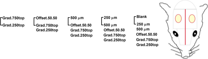
Five different calcium phosphate (CaP) coated melt electrowritten PCL pore sized scaffolds: 250 μm and 500 μm, 500 μm with 50% fibre offset (offset.50.50), tri layer gradient 250-500-750 μm (grad.250top) and 750-500-250 μm (grad.750top) were implanted into rodent critical-sized calvarial defects. Empty defects were used as a control. After 4 and 8 weeks of healing, the new bone was assessed by micro-computed tomography and immunohistochemistry.

RESULTS

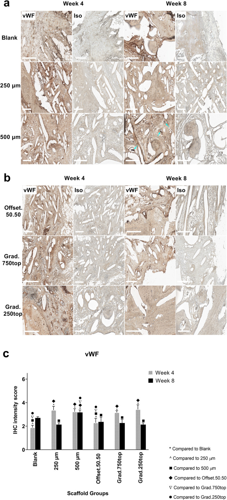
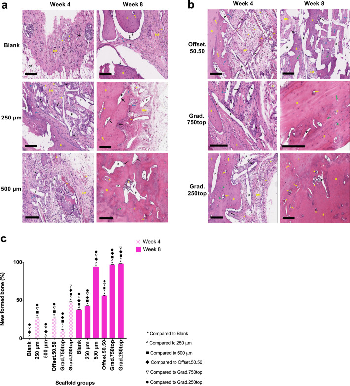
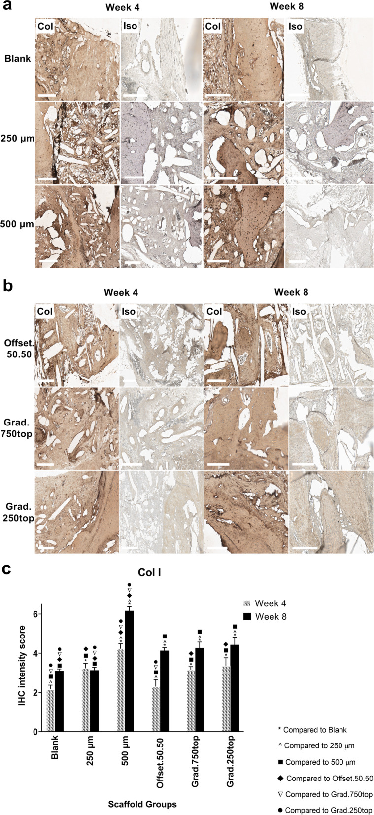
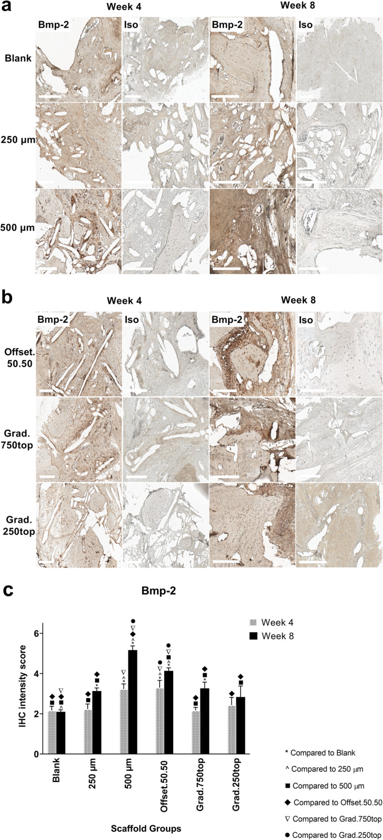
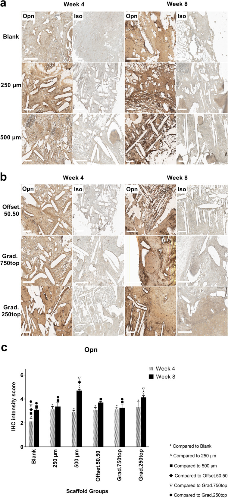
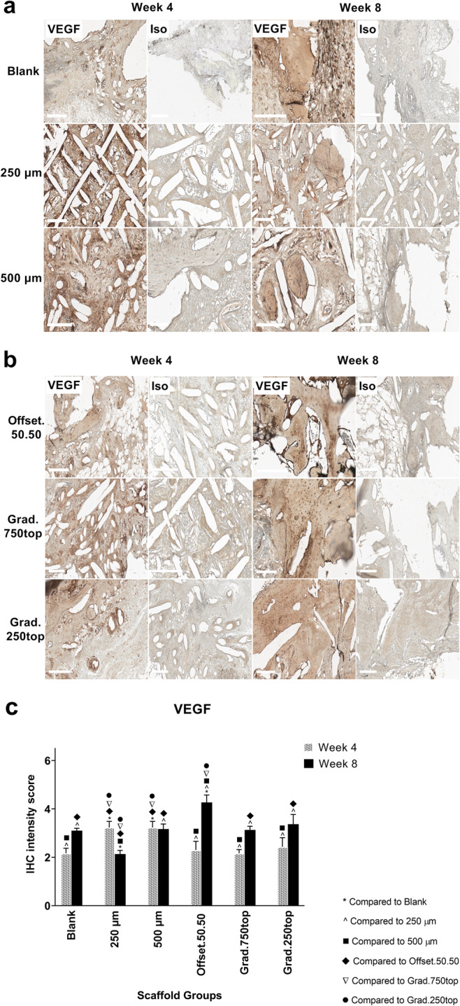
Significantly more newly formed bone was shown in the grad.250top scaffold 8 weeks post-implantation. Histological investigation also showed that soft tissue was replaced with newly formed bone and fully covered the grad.250top scaffold. While, the bone healing did not happen completely in the 250 μm, offset.50.50 scaffolds and blank calvaria defects following 8 weeks of implantation. Immunohistochemical analysis showed the expression of osteogenic markers was present in all scaffold groups at both time points. The mineralization marker Osteocalcin was detected with the highest intensity in the grad.250top and 500 μm scaffolds. Moreover, the expression of the endothelial markers showed that robust angiogenesis was involved in the repair process.

CONCLUSIONS

These results suggest that the gradient pore size structure provides superior conditions for bone regeneration.

摘要

背景

基于生物材料的骨组织工程是克服残余骨量减少的一种有前景的解决方案。先前已经证明,三维熔融电写聚己内酯(PCL)支架的梯度和偏移结构能够成功引导成骨细胞向成骨谱系分化,从而实现矿化。因此,本研究的目的是评估具有这些不同结构的PCL支架在体内的骨传导能力。

方法

将五种不同的磷酸钙(CaP)涂层熔融电写PCL孔径支架:250μm和500μm、500μm且纤维偏移50%(offset.50.50)、三层梯度250 - 500 - 750μm(grad.250top)和750 - 500 - 250μm(grad.750top)植入啮齿动物临界大小的颅骨缺损处。将空白缺损用作对照。在愈合4周和8周后,通过微计算机断层扫描和免疫组织化学评估新骨情况。

结果

植入8周后,grad.250top支架中显示出明显更多的新形成骨。组织学研究还表明,软组织被新形成的骨替代并完全覆盖了grad.250top支架。而在植入8周后,250μm、offset.50.50支架和空白颅骨缺损处的骨愈合未完全发生。免疫组织化学分析表明,在两个时间点所有支架组中均存在成骨标志物的表达。在grad.250top和500μm支架中检测到矿化标志物骨钙素的强度最高。此外,内皮标志物的表达表明强大的血管生成参与了修复过程。

结论

这些结果表明,梯度孔径结构为骨再生提供了优越条件。

https://cdn.ncbi.nlm.nih.gov/pmc/blobs/0893/7529514/b7a1940939ba/40824_2020_196_Fig15_HTML.jpg
https://cdn.ncbi.nlm.nih.gov/pmc/blobs/0893/7529514/1808a3b4242d/40824_2020_196_Fig1_HTML.jpg
https://cdn.ncbi.nlm.nih.gov/pmc/blobs/0893/7529514/98b8fce2e3d6/40824_2020_196_Fig2_HTML.jpg
https://cdn.ncbi.nlm.nih.gov/pmc/blobs/0893/7529514/b72d2b185bcc/40824_2020_196_Fig3_HTML.jpg
https://cdn.ncbi.nlm.nih.gov/pmc/blobs/0893/7529514/9a899cc907af/40824_2020_196_Fig4_HTML.jpg
https://cdn.ncbi.nlm.nih.gov/pmc/blobs/0893/7529514/dd2c50bfe582/40824_2020_196_Fig5_HTML.jpg
https://cdn.ncbi.nlm.nih.gov/pmc/blobs/0893/7529514/b72dc40548bd/40824_2020_196_Fig6_HTML.jpg
https://cdn.ncbi.nlm.nih.gov/pmc/blobs/0893/7529514/f9c988681710/40824_2020_196_Fig7_HTML.jpg
https://cdn.ncbi.nlm.nih.gov/pmc/blobs/0893/7529514/078ad8b845e5/40824_2020_196_Fig8_HTML.jpg
https://cdn.ncbi.nlm.nih.gov/pmc/blobs/0893/7529514/c73a3fbc3058/40824_2020_196_Fig9_HTML.jpg
https://cdn.ncbi.nlm.nih.gov/pmc/blobs/0893/7529514/f3fed726c8a3/40824_2020_196_Fig10_HTML.jpg
https://cdn.ncbi.nlm.nih.gov/pmc/blobs/0893/7529514/24e76eb78355/40824_2020_196_Fig11_HTML.jpg
https://cdn.ncbi.nlm.nih.gov/pmc/blobs/0893/7529514/ab0160890864/40824_2020_196_Fig12_HTML.jpg
https://cdn.ncbi.nlm.nih.gov/pmc/blobs/0893/7529514/7df18ab102e9/40824_2020_196_Fig13_HTML.jpg
https://cdn.ncbi.nlm.nih.gov/pmc/blobs/0893/7529514/4174a7e82e2f/40824_2020_196_Fig14_HTML.jpg
https://cdn.ncbi.nlm.nih.gov/pmc/blobs/0893/7529514/b7a1940939ba/40824_2020_196_Fig15_HTML.jpg

相似文献

1
In vivo bone regeneration assessment of offset and gradient melt electrowritten (MEW) PCL scaffolds.偏移和梯度熔体电写(MEW)聚己内酯(PCL)支架的体内骨再生评估
Biomater Res. 2020 Oct 1;24:17. doi: 10.1186/s40824-020-00196-1. eCollection 2020.
2
Role of offset and gradient architectures of 3-D melt electrowritten scaffold on differentiation and mineralization of osteoblasts.三维熔体电写支架的偏移和梯度结构对成骨细胞分化和矿化的作用。
Biomater Res. 2020 Jan 3;24:2. doi: 10.1186/s40824-019-0180-z. eCollection 2020.
3
Effects of Gradient and Offset Architectures on the Mechanical and Biological Properties of 3-D Melt Electrowritten (MEW) Scaffolds.梯度和偏移结构对三维熔体电写(MEW)支架的力学和生物学性能的影响
ACS Biomater Sci Eng. 2019 Jul 8;5(7):3448-3461. doi: 10.1021/acsbiomaterials.8b01456. Epub 2019 Jun 18.
4
Melt Electrowritten Sandwich Scaffold Technique Using Sulforhodamine B to Monitor Stem Cell Behavior.利用磺基罗丹明 B 通过熔融电写入三明治支架技术监测干细胞行为。
Tissue Eng Part C Methods. 2020 Oct;26(10):519-527. doi: 10.1089/ten.TEC.2020.0240.
5
The effect of pore size within fibrous scaffolds fabricated using melt electrowriting on human bone marrow stem cell osteogenesis.熔融静电纺丝制备的纤维支架中孔径对人骨髓间充质干细胞成骨的影响。
Biomed Mater. 2019 Nov 8;14(6):065016. doi: 10.1088/1748-605X/ab49f2.
6
Melt electrowriting scaffolds with fibre-guiding features for periodontal attachment.熔融静电纺丝支架具有引导纤维的特性,可用于牙周附着。
Acta Biomater. 2024 May;180:337-357. doi: 10.1016/j.actbio.2024.04.006. Epub 2024 Apr 5.
7
Assessment of bone regeneration of a tissue-engineered bone complex using human dental pulp stem cells/poly(ε-caprolactone)-biphasic calcium phosphate scaffold constructs in rabbit calvarial defects.使用人牙髓干细胞/聚(ε-己内酯)-双相磷酸钙支架构建物评估兔颅骨缺损中组织工程骨复合物的骨再生情况。
J Mater Sci Mater Med. 2017 May;28(5):77. doi: 10.1007/s10856-017-5883-x. Epub 2017 Apr 6.
8
Effect of Pore Size on Cell Behavior Using Melt Electrowritten Scaffolds.孔径对使用熔喷电写支架的细胞行为的影响。
Front Bioeng Biotechnol. 2021 Jul 2;9:629270. doi: 10.3389/fbioe.2021.629270. eCollection 2021.
9
Three-Dimensional Melt-Electrowritten Polycaprolactone/Chitosan Scaffolds Enhance Mesenchymal Stem Cell Behavior.三维熔融静电纺丝聚己内酯/壳聚糖支架增强间充质干细胞行为。
ACS Appl Bio Mater. 2021 Feb 15;4(2):1319-1329. doi: 10.1021/acsabm.0c01213. Epub 2021 Jan 13.
10
Scaffold microarchitecture regulates angiogenesis and the regeneration of large bone defects.支架的微观结构调节血管生成和大骨缺损的再生。
Biofabrication. 2022 Aug 31;14(4). doi: 10.1088/1758-5090/ac88a1.

引用本文的文献

1
Advancements in Musculoskeletal Tissue Engineering: The Role of Melt Electrowriting in 3D-Printed Scaffold Fabrication.肌肉骨骼组织工程的进展:熔体静电纺丝在3D打印支架制造中的作用。
J Funct Biomater. 2025 May 7;16(5):163. doi: 10.3390/jfb16050163.
2
Designing Smartly: Understanding the Crystallinity of Melt Electrowritten Scaffolds.巧妙设计:理解熔体电写支架的结晶度
Eng Life Sci. 2025 Apr 14;25(4):e70020. doi: 10.1002/elsc.70020. eCollection 2025 Apr.
3
Niche-inspired collagen infused melt electrowritten scaffolds for craniofacial bone regeneration.

本文引用的文献

1
Effects of Gradient and Offset Architectures on the Mechanical and Biological Properties of 3-D Melt Electrowritten (MEW) Scaffolds.梯度和偏移结构对三维熔体电写(MEW)支架的力学和生物学性能的影响
ACS Biomater Sci Eng. 2019 Jul 8;5(7):3448-3461. doi: 10.1021/acsbiomaterials.8b01456. Epub 2019 Jun 18.
2
pre-vascularization strategies for tissue engineered constructs-Bioprinting and others.组织工程构建体的血管化前策略——生物打印及其他
Int J Bioprint. 2017 Jan 23;3(1):008. doi: 10.18063/IIB.2017.01.008. eCollection 2017.
3
Role of offset and gradient architectures of 3-D melt electrowritten scaffold on differentiation and mineralization of osteoblasts.
用于颅面骨再生的受小生境启发的胶原蛋白注入式熔体电写支架
Biomater Adv. 2025 May;170:214222. doi: 10.1016/j.bioadv.2025.214222. Epub 2025 Feb 6.
4
Enhancing craniofacial bone tissue engineering strategy: integrating rapid wet chemically synthesised bioactive glass with photopolymerized resins.增强颅面骨组织工程策略:将快速湿化学合成的生物活性玻璃与光聚合树脂相结合。
BMC Oral Health. 2024 Oct 8;24(1):1195. doi: 10.1186/s12903-024-04978-0.
5
Integrating Melt Electrowriting and Fused Deposition Modeling to Fabricate Hybrid Scaffolds Supportive of Accelerated Bone Regeneration.整合熔体静电纺丝和熔融沉积建模以制造支持加速骨再生的混合支架。
Adv Healthc Mater. 2024 Jan;13(3):e2302057. doi: 10.1002/adhm.202302057. Epub 2023 Nov 16.
6
ZIF-8 induced hydroxyapatite-like crystals enabled superior osteogenic ability of MEW printing PCL scaffolds.ZIF-8 诱导生成类羟基磷灰石晶体赋予了 MEW 打印 PCL 支架优异的成骨能力。
J Nanobiotechnology. 2023 Aug 10;21(1):264. doi: 10.1186/s12951-023-02007-w.
7
FLIM imaging revealed spontaneous osteogenic differentiation of stem cells on gradient pore size tissue-engineered constructs.荧光寿命成像显示,干细胞在梯度孔径组织工程构建体上自发地向成骨细胞分化。
Stem Cell Res Ther. 2023 Apr 12;14(1):81. doi: 10.1186/s13287-023-03307-6.
8
Double-Reinforced Fish Gelatin Composite Scaffolds for Osteochondral Substitutes.用于骨软骨替代物的双增强鱼明胶复合支架
Materials (Basel). 2023 Feb 22;16(5):1815. doi: 10.3390/ma16051815.
9
Advanced biomaterials for periodontal tissue regeneration.牙周组织再生的先进生物材料。
Genesis. 2022 Sep;60(8-9):e23501. doi: 10.1002/dvg.23501. Epub 2022 Sep 16.
10
Recent advances in melt electro writing for tissue engineering for 3D printing of microporous scaffolds for tissue engineering.用于组织工程的微孔支架3D打印的熔体电写技术在组织工程领域的最新进展。
Front Bioeng Biotechnol. 2022 Aug 17;10:896719. doi: 10.3389/fbioe.2022.896719. eCollection 2022.
三维熔体电写支架的偏移和梯度结构对成骨细胞分化和矿化的作用。
Biomater Res. 2020 Jan 3;24:2. doi: 10.1186/s40824-019-0180-z. eCollection 2020.
4
Calvarial Versus Long Bone: Implications for Tailoring Skeletal Tissue Engineering.颅骨与长骨:对骨骼组织工程定制的启示。
Tissue Eng Part B Rev. 2020 Feb;26(1):46-63. doi: 10.1089/ten.TEB.2018.0353. Epub 2019 Dec 18.
5
A Novel Bilayer Polycaprolactone Membrane for Guided Bone Regeneration: Combining Electrospinning and Emulsion Templating.一种用于引导骨再生的新型双层聚己内酯膜:静电纺丝与乳液模板法相结合
Materials (Basel). 2019 Aug 20;12(16):2643. doi: 10.3390/ma12162643.
6
Integrated Design Approaches for 3D Printed Tissue Scaffolds: Review and Outlook.3D打印组织支架的集成设计方法:综述与展望
Materials (Basel). 2019 Jul 24;12(15):2355. doi: 10.3390/ma12152355.
7
Synthesis and Evaluation of BMMSC-seeded BMP-6/nHAG/GMS Scaffolds for Bone Regeneration.骨髓间充质干细胞负载 BMP-6/纳米羟基磷灰石/明胶支架的制备及其对骨再生的评价
Int J Med Sci. 2019 Jun 10;16(7):1007-1017. doi: 10.7150/ijms.31966. eCollection 2019.
8
Strategies to Tune Electrospun Scaffold Porosity for Effective Cell Response in Tissue Engineering.调控静电纺丝支架孔隙率以实现组织工程中有效细胞响应的策略
J Funct Biomater. 2019 Jul 9;10(3):30. doi: 10.3390/jfb10030030.
9
The Applications of 3D Printing for Craniofacial Tissue Engineering.3D打印在颅面组织工程中的应用
Micromachines (Basel). 2019 Jul 17;10(7):480. doi: 10.3390/mi10070480.
10
Enhanced bone tissue regeneration of a biomimetic cellular scaffold with co-cultured MSCs-derived osteogenic and angiogenic cells.仿生细胞支架共培养 MSC 源性成骨和成血管细胞增强骨组织再生。
Cell Prolif. 2019 Sep;52(5):e12658. doi: 10.1111/cpr.12658. Epub 2019 Jul 11.