Shakya Reshma, Nguyen Tam Hong, Waterhouse Nigel, Khanna Rajiv
QIMR Berghofer Centre for Immunotherapy and Vaccine Development, Tumour Immunology Laboratory QIMR Berghofer Medical Research Institute Brisbane QLD Australia.
Flow Cytometry and Imaging Facility QIMR Berghofer Medical Research Institute Brisbane QLD Australia.
Clin Transl Immunology. 2020 Oct 7;9(10):e1183. doi: 10.1002/cti2.1183. eCollection 2020.
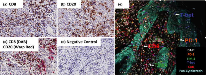
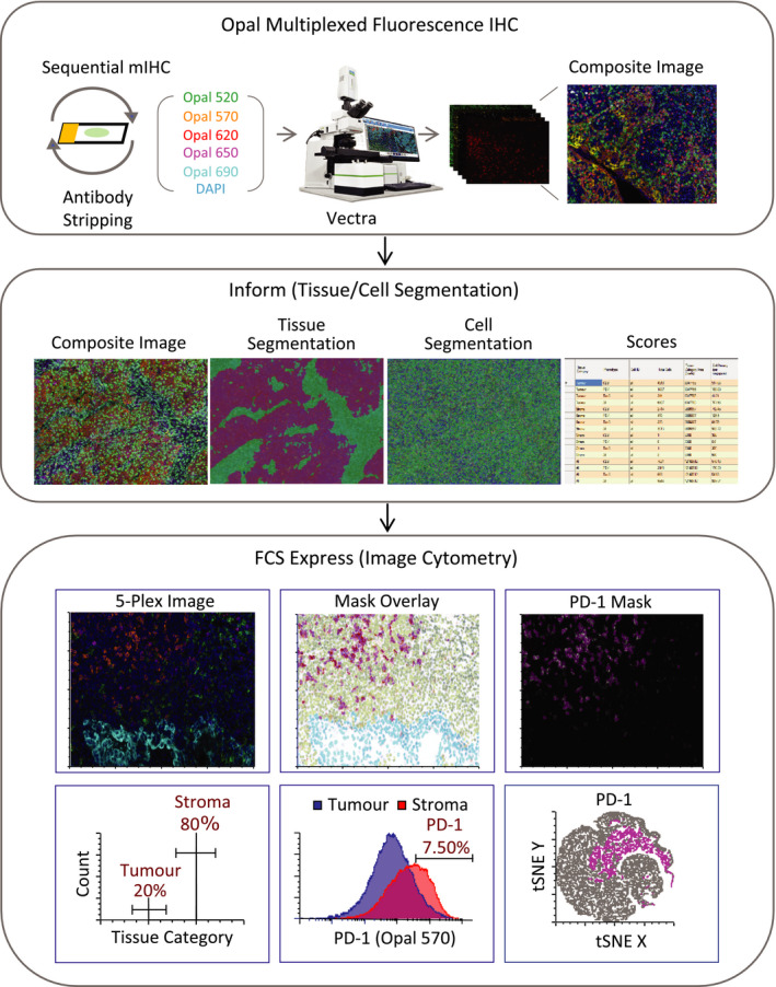
The tumor microenvironment is an integral player in cancer initiation, tumor progression, response and resistance to anti-cancer therapy. Understanding the complex interactions of tumor immune architecture (referred to as 'immune contexture') has therefore become increasingly desirable to guide our approach to patient selection, clinical trial design, combination therapies, and patient management. Quantitative image analysis based on multiplexed fluorescence immunohistochemistry and deep learning technologies are rapidly developing to enable researchers to interrogate complex information from the tumor microenvironment and find predictive insights into treatment response. Herein, we discuss current developments in multiplexed fluorescence immunohistochemistry for immune contexture analysis, and their application in immuno-oncology, and discuss challenges to effectively use this technology in clinical settings. We also present a multiplexed image analysis workflow to analyse fluorescence multiplexed stained tumor sections using the Vectra Automated Digital Pathology System together with FCS express flow cytometry software. The benefit of this strategy is that the spectral unmixing accurately generates and analyses complex arrays of multiple biomarkers, which can be helpful for diagnosis, risk stratification, and guiding clinical management of oncology patients.
肿瘤微环境在癌症的发生、肿瘤进展、对抗癌治疗的反应及耐药性方面发挥着不可或缺的作用。因此,了解肿瘤免疫结构(称为“免疫格局”)的复杂相互作用,对于指导我们进行患者选择、临床试验设计、联合治疗及患者管理而言,变得越来越必要。基于多重荧光免疫组织化学和深度学习技术的定量图像分析正在迅速发展,使研究人员能够从肿瘤微环境中探究复杂信息,并找到有关治疗反应的预测性见解。在此,我们讨论用于免疫格局分析的多重荧光免疫组织化学的当前进展及其在免疫肿瘤学中的应用,并讨论在临床环境中有效使用该技术所面临的挑战。我们还展示了一种多重图像分析工作流程,以使用Vectra自动数字病理系统和FCS express流式细胞术软件分析荧光多重染色的肿瘤切片。该策略的优势在于光谱解混能够准确生成并分析多种生物标志物的复杂阵列,这有助于肿瘤患者的诊断、风险分层及临床管理指导。