Mastropasqua Rodolfo, Evangelista Federica, Amodei Francesco, D'Aloisio Rossella, Pinto Filomena, Doronzo Emanuele, Viggiano Pasquale, Porreca Annamaria, Di Nicola Marta, Parravano Mariacristina, Toto Lisa
Institute of Ophthalmology, University of Modena and Reggio Emilia, Modena, Italy.
Ophthalmic Clinic, Department of Medicine and Science of Ageing, University G. D'Annunzio Chieti-Pescara, Chieti, Italy.
Transl Vis Sci Technol. 2020 Oct 7;9(11):6. doi: 10.1167/tvst.9.11.6. eCollection 2020 Oct.
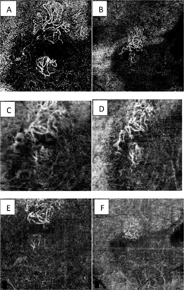
The purpose of this study is to compare the ability of 3 optical coherence tomography angiography (OCTA) devices to measure lesion area in patients with macular neovascularization (MNV) with type 1, 2 and mixed neovascularization (NV).
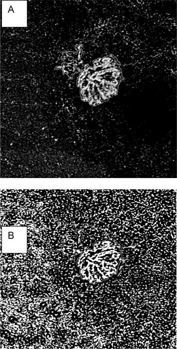
OCTA, fluorescein angiography (FA), indocyanine green angiography (ICGA), and structural optical coherence tomography (OCT) were performed. NV lesion area measurements were performed by two graders.
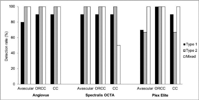
Twenty-eight eyes were included: 20 with NV were classified as type 1, 6 as type 2, and 2 as mixed type. AngioVue and Spectralis detected the NV in 26 out of 28 eyes (92.8%). The intraclass correlation coefficient (ICC) between readers for the three different OCTA with the different slabs was high. The NV area was larger in the outer retina to choriocapillaris (ORCC) and choriocapillaris (CC) images for the AngioVue device and the PLEX Elite device compared to avascular images ( < 0.05). The mean values of the NV area were not significantly different among the three instruments (Friedman test, > 0.05) for the avascular zone (AV), ORCC, and CC images. Median (interquartile range [IQR]) NV were significantly different among avascular images, ORCC images, and CC images of the AngioVue device ( = 0.046), of the Spectralis device ( = 0.015), and the PLEX Elite device ( < 0.001).
The ORCC slabs showed the highest detection rate for NV detection independently to the device used, and swept source (SS)-OCTA measurements of ORCC slabs showed the highest detection rate of NVs compared to the spectral domain (SD)-OCTA.
It is pivotal to realize how much we can rely on OCTA to make a diagnosis of NV.
本研究旨在比较3种光学相干断层扫描血管造影(OCTA)设备测量1型、2型及混合型黄斑新生血管(MNV)患者病变面积的能力。
进行了OCTA、荧光素血管造影(FA)、吲哚菁绿血管造影(ICGA)和结构光学相干断层扫描(OCT)检查。由两名分级者进行新生血管病变面积测量。
纳入28只眼:20只新生血管眼分类为1型,6只为2型,2只为混合型。AngioVue和Spectralis在28只眼中的26只(92.8%)检测到了新生血管。不同平板的三种不同OCTA在阅片者之间的组内相关系数(ICC)较高。与无血管图像相比,AngioVue设备和PLEX Elite设备在外视网膜至脉络膜毛细血管(ORCC)和脉络膜毛细血管(CC)图像中的新生血管面积更大(P<0.05)。对于无血管区(AV)、ORCC和CC图像,三种仪器之间新生血管面积的平均值无显著差异(Friedman检验,P>0.05)。AngioVue设备、Spectralis设备和PLEX Elite设备的无血管图像、ORCC图像和CC图像中的新生血管中位数(四分位间距[IQR])有显著差异(分别为P=0.046、P=0.015和P<0.001)。
无论使用何种设备,ORCC平板对新生血管的检测率最高,与谱域(SD)-OCTA相比,扫频源(SS)-OCTA对ORCC平板的新生血管检测率最高。
了解我们在多大程度上可以依靠OCTA来诊断新生血管至关重要。