Lin Wenjiao, Zhang Hongjie, Zhang Wanqian, Qi Haiping, Zhang Gui, Qian Jie, Li Xin, Qin Li, Li Haifeng, Wang Xiang, Qiu Hong, Shi Xiaoli, Zheng Wei, Zhang Deyuan, Gao Runlin, Ding Jiandong
College of Materials Science and Chemical Engineering, Harbin Engineering University, Harbin 150001, China.
Biotyx Medical (Shenzhen) Co., Ltd, Shenzhen 518109, China.
Bioact Mater. 2020 Oct 12;6(4):1028-1039. doi: 10.1016/j.bioactmat.2020.09.020. eCollection 2021 Apr.
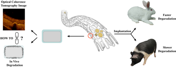
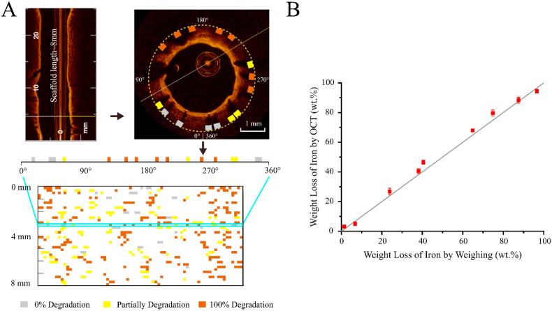
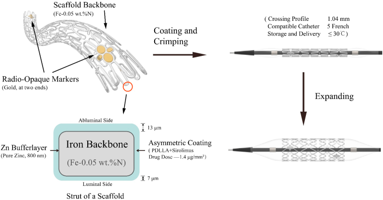
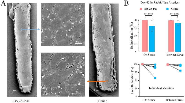
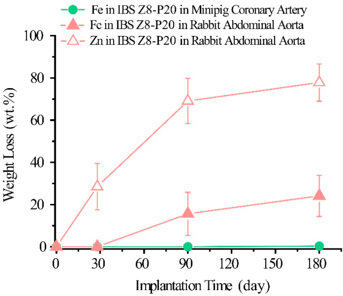
Detection of in vivo biodegradation is critical for development of next-generation medical devices such as bioresorbable stents or scaffolds (BRSs). In particular, it is urgent to establish a nondestructive approach to examine in vivo degradation of a new-generation coronary stent for interventional treatment based on mammal experiments; otherwise it is not available to semi-quantitatively monitor biodegradation in any clinical trial. Herein, we put forward a semi-quantitative approach to measure degradation of a sirolimus-eluting iron bioresorbable scaffold (IBS) based on optical coherence tomography (OCT) images; this approach was confirmed to be consistent with the present weight-loss measurements, which is, however, a destructive approach. The IBS was fabricated by a metal-polymer composite technique with a polylactide coating on an iron stent. The efficacy as a coronary stent of this new bioresorbable scaffold was compared with that of a permanent metal stent with the name of trade mark Xience, which has been widely used in clinic. The endothelial coverage on IBS was found to be greater than on Xience after implantation in a rabbit model; and our well-designed ultrathin stent exhibited less individual variation. We further examined degradation of the IBSs in both minipig coronary artery and rabbit abdominal aorta models. The present result indicated much faster iron degradation of IBS in the rabbit model than in the porcine model. The semi-quantitative approach to detect biodegradation of IBS and the finding of the species difference might be stimulating for fundamental investigation of biodegradable implants and clinical translation of the next-generation coronary stents.
检测体内生物降解对于下一代医疗设备(如生物可吸收支架或支架)的开发至关重要。特别是,迫切需要建立一种非破坏性方法,以基于哺乳动物实验来检查用于介入治疗的新一代冠状动脉支架的体内降解情况;否则,在任何临床试验中都无法对生物降解进行半定量监测。在此,我们提出了一种基于光学相干断层扫描(OCT)图像来测量西罗莫司洗脱铁生物可吸收支架(IBS)降解的半定量方法;该方法被证实与目前的失重测量结果一致,然而,失重测量是一种破坏性方法。IBS是通过金属 - 聚合物复合技术在铁支架上涂覆聚丙交酯制成的。将这种新型生物可吸收支架作为冠状动脉支架的疗效与已在临床上广泛使用的名为Xience的永久性金属支架进行了比较。在兔模型中植入后发现,IBS上的内皮覆盖率高于Xience;并且我们精心设计的超薄支架个体差异较小。我们进一步在小型猪冠状动脉和兔腹主动脉模型中检查了IBS的降解情况。目前的结果表明,IBS在兔模型中的铁降解比在猪模型中快得多。检测IBS生物降解的半定量方法以及物种差异的发现可能会刺激对可生物降解植入物的基础研究以及下一代冠状动脉支架的临床转化。