Center for Brain Immunology and Glia, Department of Neuroscience, University of Virginia, Charlottesville, Virginia, United States of America.
Baker Institute for Animal Health and Department of Microbiology and Immunology, Cornell University, Ithaca, New York, United States of America.
PLoS Pathog. 2020 Oct 27;16(10):e1009027. doi: 10.1371/journal.ppat.1009027. eCollection 2020 Oct.
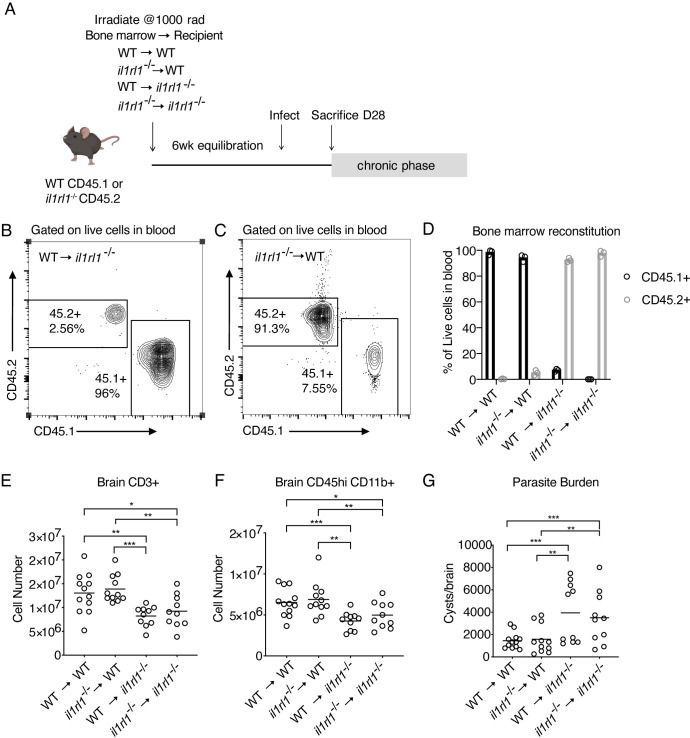
It is of great interest to understand how invading pathogens are sensed within the brain, a tissue with unique challenges to mounting an immune response. The eukaryotic parasite Toxoplasma gondii colonizes the brain of its hosts, and initiates robust immune cell recruitment, but little is known about pattern recognition of T. gondii within brain tissue. The host damage signal IL-33 is one protein that has been implicated in control of chronic T. gondii infection, but, like many other pattern recognition pathways, IL-33 can signal peripherally, and the specific impact of IL-33 signaling within the brain is unclear. Here, we show that IL-33 is expressed by oligodendrocytes and astrocytes during T. gondii infection, is released locally into the cerebrospinal fluid of T. gondii-infected animals, and is required for control of infection. IL-33 signaling promotes chemokine expression within brain tissue and is required for the recruitment and/or maintenance of blood-derived anti-parasitic immune cells, including proliferating, IFN-γ-expressing T cells and iNOS-expressing monocytes. Importantly, we find that the beneficial effects of IL-33 during chronic infection are not a result of signaling on infiltrating immune cells, but rather on radio-resistant responders, and specifically, astrocytes. Mice with IL-33 receptor-deficient astrocytes fail to mount an adequate adaptive immune response in the CNS to control parasite burden-demonstrating, genetically, that astrocytes can directly respond to IL-33 in vivo. Together, these results indicate a brain-specific mechanism by which IL-33 is released locally, and sensed locally, to engage the peripheral immune system in controlling a pathogen.
了解入侵病原体如何在大脑中被感知是非常重要的,因为大脑组织在引发免疫反应方面具有独特的挑战。真核寄生虫刚地弓形虫定植在宿主的大脑中,并引发强烈的免疫细胞募集,但对于大脑组织中刚地弓形虫的模式识别知之甚少。宿主损伤信号 IL-33 是一种被认为可以控制慢性刚地弓形虫感染的蛋白质,但与许多其他模式识别途径一样,IL-33 可以在周围组织中信号传递,IL-33 信号在大脑内的具体影响尚不清楚。在这里,我们表明,IL-33 在刚地弓形虫感染期间由少突胶质细胞和星形胶质细胞表达,局部释放到刚地弓形虫感染动物的脑脊液中,并控制感染。IL-33 信号促进脑组织中趋化因子的表达,并且对于招募和/或维持血液来源的抗寄生虫免疫细胞是必需的,包括增殖的 IFN-γ 表达 T 细胞和 iNOS 表达单核细胞。重要的是,我们发现 IL-33 在慢性感染期间的有益作用不是由于浸润免疫细胞的信号传递,而是由于对辐射抗性反应者,特别是星形胶质细胞的信号传递。缺乏 IL-33 受体的星形胶质细胞的小鼠不能在中枢神经系统中产生足够的适应性免疫反应来控制寄生虫负担-从基因上证明了星形胶质细胞可以在体内直接对 IL-33 作出反应。总之,这些结果表明了一种大脑特异性机制,通过该机制,IL-33 局部释放并局部感知,从而使外周免疫系统参与控制病原体。