• 文献检索
  • 文档翻译
  • 深度研究
  • 学术资讯
  • Suppr Zotero 插件Zotero 插件
  • 邀请有礼
  • 套餐&价格
  • 历史记录
应用&插件
Suppr Zotero 插件Zotero 插件浏览器插件Mac 客户端Windows 客户端微信小程序
定价
高级版会员购买积分包购买API积分包
服务
文献检索文档翻译深度研究API 文档MCP 服务
关于我们
关于 Suppr公司介绍联系我们用户协议隐私条款
关注我们

Suppr 超能文献

核心技术专利:CN118964589B侵权必究
粤ICP备2023148730 号-1Suppr @ 2026

文献检索

告别复杂PubMed语法,用中文像聊天一样搜索,搜遍4000万医学文献。AI智能推荐,让科研检索更轻松。

立即免费搜索

文件翻译

保留排版,准确专业,支持PDF/Word/PPT等文件格式,支持 12+语言互译。

免费翻译文档

深度研究

AI帮你快速写综述,25分钟生成高质量综述,智能提取关键信息,辅助科研写作。

立即免费体验

磁共振影像组学分析能否鉴别腮腺肿瘤?一项初步研究。

Can Magnetic Resonance Radiomics Analysis Discriminate Parotid Gland Tumors? A Pilot Study.

作者信息

Gabelloni Michela, Faggioni Lorenzo, Attanasio Simona, Vani Vanina, Goddi Antonio, Colantonio Sara, Germanese Danila, Caudai Claudia, Bruschini Luca, Scarano Mariella, Seccia Veronica, Neri Emanuele

机构信息

Diagnostic and Interventional Radiology, Department of Translational Research, University of Pisa, Via Roma, 67, 56126 Pisa, Italy.

Institute of Information Science and Technologies "A. Faedo" of the National Research Council of Italy (ISTI-CNR), 56124 Pisa, Italy.

出版信息

Diagnostics (Basel). 2020 Nov 3;10(11):900. doi: 10.3390/diagnostics10110900.

DOI:10.3390/diagnostics10110900
PMID:33153140
原文链接:https://pmc.ncbi.nlm.nih.gov/articles/PMC7692594/
Abstract

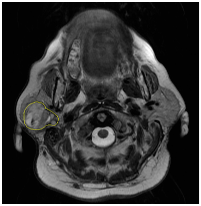
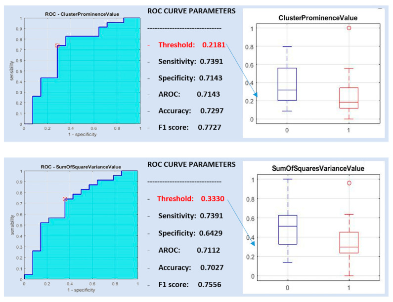
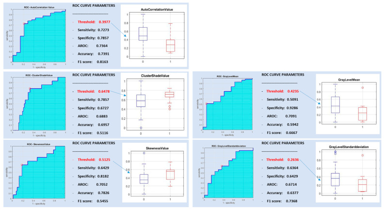
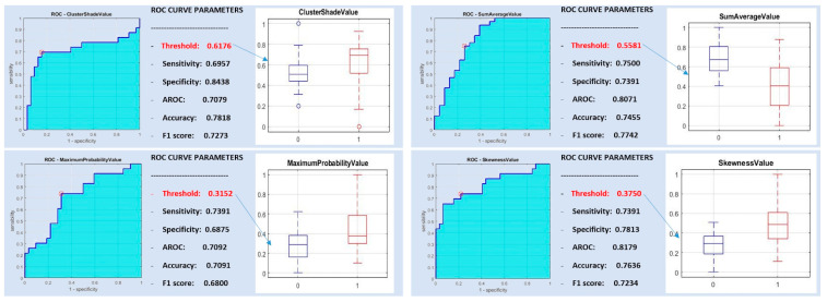
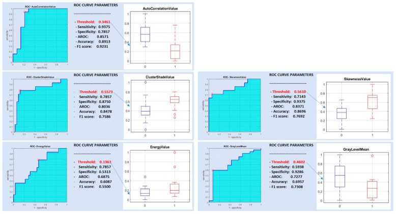
Our purpose is to evaluate the performance of magnetic resonance (MR) radiomics analysis for differentiating between malignant and benign parotid neoplasms and, among the latter, between pleomorphic adenomas and Warthin tumors. We retrospectively evaluated 75 T2-weighted images of parotid gland lesions, of which 61 were benign tumors (32 pleomorphic adenomas, 23 Warthin tumors and 6 oncocytomas) and 14 were malignant tumors. A receiver operating characteristics (ROC) curve analysis was performed to find the threshold values for the most discriminative features and determine their sensitivity, specificity and area under the ROC curve (AUROC). The most discriminative features were used to train a support vector machine classifier. The best classification performance was obtained by comparing a pleomorphic adenoma with a Warthin tumor (yielding sensitivity, specificity and a diagnostic accuracy as high as 0.8695, 0.9062 and 0.8909, respectively) and a pleomorphic adenoma with malignant tumors (sensitivity, specificity and a diagnostic accuracy of 0.6666, 0.8709 and 0.8043, respectively). Radiomics analysis of parotid tumors on conventional T2-weighted MR images allows the discrimination of pleomorphic adenomas from Warthin tumors and malignant tumors with a high sensitivity, specificity and diagnostic accuracy.

摘要

我们的目的是评估磁共振(MR)影像组学分析在鉴别腮腺良恶性肿瘤以及在良性肿瘤中鉴别多形性腺瘤和沃辛瘤的性能。我们回顾性评估了75例腮腺病变的T2加权图像,其中61例为良性肿瘤(32例多形性腺瘤、23例沃辛瘤和6例嗜酸细胞瘤),14例为恶性肿瘤。进行了受试者操作特征(ROC)曲线分析,以找出最具鉴别力特征的阈值,并确定其敏感性、特异性和ROC曲线下面积(AUROC)。使用最具鉴别力的特征训练支持向量机分类器。通过比较多形性腺瘤与沃辛瘤(敏感性、特异性和诊断准确性分别高达0.8695、0.9062和0.8909)以及多形性腺瘤与恶性肿瘤(敏感性、特异性和诊断准确性分别为0.6666、0.8709和0.8043),获得了最佳分类性能。对常规T2加权MR图像上的腮腺肿瘤进行影像组学分析,能够以较高的敏感性、特异性和诊断准确性鉴别多形性腺瘤与沃辛瘤以及恶性肿瘤。

https://cdn.ncbi.nlm.nih.gov/pmc/blobs/63ab/7692594/60dfd1d25d00/diagnostics-10-00900-g005.jpg
https://cdn.ncbi.nlm.nih.gov/pmc/blobs/63ab/7692594/bc35d632957f/diagnostics-10-00900-g001.jpg
https://cdn.ncbi.nlm.nih.gov/pmc/blobs/63ab/7692594/246ebd14ae7f/diagnostics-10-00900-g002.jpg
https://cdn.ncbi.nlm.nih.gov/pmc/blobs/63ab/7692594/d41c3f576cf3/diagnostics-10-00900-g003.jpg
https://cdn.ncbi.nlm.nih.gov/pmc/blobs/63ab/7692594/6d1cc2d0eff4/diagnostics-10-00900-g004.jpg
https://cdn.ncbi.nlm.nih.gov/pmc/blobs/63ab/7692594/60dfd1d25d00/diagnostics-10-00900-g005.jpg

相似文献

1
Can Magnetic Resonance Radiomics Analysis Discriminate Parotid Gland Tumors? A Pilot Study.磁共振影像组学分析能否鉴别腮腺肿瘤?一项初步研究。
Diagnostics (Basel). 2020 Nov 3;10(11):900. doi: 10.3390/diagnostics10110900.
2
Diagnostic performance of qualitative and radiomics approach to parotid gland tumors: which is the added benefit of texture analysis?定性和影像组学方法诊断腮腺肿瘤的性能:纹理分析的附加益处是什么?
Br J Radiol. 2021 Dec;94(1128):20210340. doi: 10.1259/bjr.20210340. Epub 2021 Sep 30.
3
Value of T2-weighted-based radiomics model in distinguishing Warthin tumor from pleomorphic adenoma of the parotid.基于 T2 加权的放射组学模型在鉴别腮腺沃辛瘤与多形性腺瘤中的价值。
Eur Radiol. 2023 Jun;33(6):4453-4463. doi: 10.1007/s00330-022-09295-0. Epub 2022 Dec 11.
4
Texture-based and diffusion-weighted discrimination of parotid gland lesions on MR images at 3.0 Tesla.基于纹理和扩散加权的 3.0T MRI 腮腺病变鉴别。
NMR Biomed. 2013 Nov;26(11):1372-9. doi: 10.1002/nbm.2962. Epub 2013 May 23.
5
Usefulness of a Novel Ultrasonographic Classification Based on Anechoic Area Patterns for Differentiating Warthin Tumors from Pleomorphic Adenomas of the Parotid Gland.基于无回声区模式的新型超声分类法在鉴别腮腺沃辛瘤与多形性腺瘤中的应用价值。
Yonago Acta Med. 2018 Feb 5;60(4):220-226. doi: 10.24563/yam.2017.12.002. eCollection 2017 Dec.
6
Usefulness of MRI-based radiomic features for distinguishing Warthin tumor from pleomorphic adenoma: performance assessment using T2-weighted and post-contrast T1-weighted MR images.基于MRI的影像组学特征在鉴别沃辛瘤与多形性腺瘤中的应用:使用T2加权和对比增强T1加权MR图像的性能评估
Eur J Radiol Open. 2022 Jun 18;9:100429. doi: 10.1016/j.ejro.2022.100429. eCollection 2022.
7
Enhanced CT-based texture analysis and radiomics score for differentiation of pleomorphic adenoma, basal cell adenoma, and Warthin tumor of the parotid gland.基于增强 CT 的纹理分析和放射组学评分在鉴别腮腺多形性腺瘤、基底细胞腺瘤和沃辛瘤中的应用。
Dentomaxillofac Radiol. 2023 Jan;52(2):20220009. doi: 10.1259/dmfr.20220009. Epub 2023 Jan 3.
8
Apparent diffusion coefficient (ADC) histogram analysis: differentiation of benign from malignant parotid gland tumors using readout-segmented diffusion-weighted imaging.表观扩散系数(ADC)直方图分析:应用分段读出扩散加权成像对腮腺良恶性肿瘤进行鉴别诊断。
Dentomaxillofac Radiol. 2019 Oct;48(7):20190100. doi: 10.1259/dmfr.20190100. Epub 2019 Jul 9.
9
Differentiation of Benign From Malignant Parotid Gland Tumors Using Conventional MRI Based on Radiomics Nomogram.基于影像组学列线图的传统MRI鉴别腮腺良恶性肿瘤
Front Oncol. 2022 Jul 11;12:937050. doi: 10.3389/fonc.2022.937050. eCollection 2022.
10
An ultrasound-based ensemble machine learning model for the preoperative classification of pleomorphic adenoma and Warthin tumor in the parotid gland.基于超声的集成机器学习模型用于术前腮腺多形性腺瘤和沃辛瘤的分类。
Eur Radiol. 2024 Oct;34(10):6862-6876. doi: 10.1007/s00330-024-10719-2. Epub 2024 Apr 3.

引用本文的文献

1
Distinguishing atypical parotid carcinomas and pleomorphic adenomas based on multiphasic computed tomography radiomics nomogram: a multicenter study.基于多期计算机断层扫描影像组学列线图鉴别非典型腮腺癌和多形性腺瘤:一项多中心研究
Front Oncol. 2025 Aug 1;15:1625487. doi: 10.3389/fonc.2025.1625487. eCollection 2025.
2
Classification of Parotid Tumors with Robust Radiomic Features from DCE- and DW-MRI.利用动态对比增强磁共振成像(DCE-MRI)和扩散加权磁共振成像(DW-MRI)的稳健影像组学特征对腮腺肿瘤进行分类
J Imaging. 2025 Apr 17;11(4):122. doi: 10.3390/jimaging11040122.
3
Nomogram combining dual-energy computed tomography features and radiomics for differentiating parotid warthin tumor from pleomorphic adenoma: a retrospective study.

本文引用的文献

1
Imaging biomarkers in upper gastrointestinal cancers.上消化道癌症中的影像生物标志物
BJR Open. 2019 Jun 24;1(1):20190001. doi: 10.1259/bjro.20190001. eCollection 2019.
2
A review of original articles published in the emerging field of radiomics.一篇关于放射组学这一新兴领域的原始文章的综述。
Eur J Radiol. 2020 Jun;127:108991. doi: 10.1016/j.ejrad.2020.108991. Epub 2020 Apr 12.
3
Tumour heterogeneity revealed by unsupervised decomposition of dynamic contrast-enhanced magnetic resonance imaging is associated with underlying gene expression patterns and poor survival in breast cancer patients.
结合双能计算机断层扫描特征和放射组学鉴别腮腺沃辛瘤与多形性腺瘤的列线图:一项回顾性研究
Front Oncol. 2025 Mar 4;15:1505385. doi: 10.3389/fonc.2025.1505385. eCollection 2025.
4
The Role of MRI Radiomics Using T2-Weighted Images and the Apparent Diffusion Coefficient Map for Discriminating Between Warthin's Tumors and Malignant Parotid Gland Tumors.利用T2加权图像和表观扩散系数图的MRI影像组学在鉴别沃辛瘤和腮腺恶性肿瘤中的作用
Cancers (Basel). 2025 Feb 12;17(4):620. doi: 10.3390/cancers17040620.
5
Assessment of synthetic MRI to distinguish Warthin's tumor from pleomorphic adenoma in the parotid gland: comparison of two methods of positioning the region of interest for synthetic relaxometry measurement.评估合成磁共振成像以鉴别腮腺沃辛瘤与多形性腺瘤:两种合成弛豫测量感兴趣区定位方法的比较
Front Oncol. 2024 Oct 4;14:1446736. doi: 10.3389/fonc.2024.1446736. eCollection 2024.
6
Using Machine Learning on MRI Radiomics to Diagnose Parotid Tumours Before Comparing Performance with Radiologists: A Pilot Study.在将性能与放射科医生进行比较之前,利用机器学习对MRI影像组学进行腮腺肿瘤诊断:一项初步研究。
J Imaging Inform Med. 2025 Jun;38(3):1496-1508. doi: 10.1007/s10278-024-01255-y. Epub 2024 Oct 10.
7
Diagnostic performance of MRI-based radiomics models using machine learning approaches for the triple classification of parotid tumors.使用机器学习方法的基于MRI的放射组学模型对腮腺肿瘤进行三重分类的诊断性能。
Heliyon. 2024 Aug 22;10(17):e36601. doi: 10.1016/j.heliyon.2024.e36601. eCollection 2024 Sep 15.
8
Prediction of malignant risk stratification model for parotid gland nodules based on clinical and conventional ultrasound features: construction and validation.基于临床和常规超声特征的腮腺结节恶性风险分层模型的预测:构建与验证
Gland Surg. 2024 Jul 30;13(7):1229-1242. doi: 10.21037/gs-24-119. Epub 2024 Jul 24.
9
The value of T1- and FST2-Weighted-based radiomics nomogram in differentiating pleomorphic adenoma and Warthin tumor.基于T1加权和FST2加权的影像组学列线图在鉴别多形性腺瘤和沃辛瘤中的价值。
Transl Oncol. 2024 Nov;49:102087. doi: 10.1016/j.tranon.2024.102087. Epub 2024 Aug 18.
10
Performance of radiomics in the differential diagnosis of parotid tumors: a systematic review.放射组学在腮腺肿瘤鉴别诊断中的应用:一项系统评价
Front Oncol. 2024 Jul 25;14:1383323. doi: 10.3389/fonc.2024.1383323. eCollection 2024.
非监督性动态对比增强磁共振成像分解所揭示的肿瘤异质性与乳腺癌患者的潜在基因表达模式和不良预后相关。
Breast Cancer Res. 2019 Oct 17;21(1):112. doi: 10.1186/s13058-019-1199-8.
4
Image-based Classification of Tumor Type and Growth Rate using Machine Learning: a preclinical study.基于图像的肿瘤类型和生长速度的机器学习分类:一项临床前研究。
Sci Rep. 2019 Aug 29;9(1):12529. doi: 10.1038/s41598-019-48738-5.
5
Proteomic Investigation of Malignant Major Salivary Gland Tumors.恶性涎腺大涎腺肿瘤的蛋白质组学研究。
Head Neck Pathol. 2020 Jun;14(2):362-373. doi: 10.1007/s12105-019-01040-2. Epub 2019 May 16.
6
Radiomics and liquid biopsy in oncology: the holons of systems medicine.肿瘤学中的放射组学与液体活检:系统医学的整体元
Insights Imaging. 2018 Dec;9(6):915-924. doi: 10.1007/s13244-018-0657-7. Epub 2018 Nov 14.
7
Association between tumor heterogeneity and progression-free survival in non-small cell lung cancer patients with EGFR mutations undergoing tyrosine kinase inhibitors therapy.接受酪氨酸激酶抑制剂治疗的表皮生长因子受体(EGFR)突变的非小细胞肺癌患者中肿瘤异质性与无进展生存期的关联。
Annu Int Conf IEEE Eng Med Biol Soc. 2016 Aug;2016:1268-1271. doi: 10.1109/EMBC.2016.7590937.
8
MRI texture analysis (MRTA) of T2-weighted images in Crohn's disease may provide information on histological and MRI disease activity in patients undergoing ileal resection.克罗恩病患者T2加权图像的MRI纹理分析(MRTA)可能为接受回肠切除术患者的组织学及MRI疾病活动提供信息。
Eur Radiol. 2017 Feb;27(2):589-597. doi: 10.1007/s00330-016-4324-4. Epub 2016 Apr 5.
9
Differential diagnosis of parotid gland tumours: which magnetic resonance findings should be taken in account?腮腺肿瘤的鉴别诊断:应考虑哪些磁共振成像结果?
Acta Otorhinolaryngol Ital. 2015 Oct;35(5):314-20. doi: 10.14639/0392-100X-693.
10
Haralick texture analysis of prostate MRI: utility for differentiating non-cancerous prostate from prostate cancer and differentiating prostate cancers with different Gleason scores.前列腺MRI的哈拉里克纹理分析:用于区分非癌性前列腺与前列腺癌以及区分不同Gleason评分的前列腺癌的效用。
Eur Radiol. 2015 Oct;25(10):2840-50. doi: 10.1007/s00330-015-3701-8. Epub 2015 May 21.