• 文献检索
  • 文档翻译
  • 深度研究
  • 学术资讯
  • Suppr Zotero 插件Zotero 插件
  • 邀请有礼
  • 套餐&价格
  • 历史记录
应用&插件
Suppr Zotero 插件Zotero 插件浏览器插件Mac 客户端Windows 客户端微信小程序
定价
高级版会员购买积分包购买API积分包
服务
文献检索文档翻译深度研究API 文档MCP 服务
关于我们
关于 Suppr公司介绍联系我们用户协议隐私条款
关注我们

Suppr 超能文献

核心技术专利:CN118964589B侵权必究
粤ICP备2023148730 号-1Suppr @ 2026

文献检索

告别复杂PubMed语法,用中文像聊天一样搜索,搜遍4000万医学文献。AI智能推荐,让科研检索更轻松。

立即免费搜索

文件翻译

保留排版,准确专业,支持PDF/Word/PPT等文件格式,支持 12+语言互译。

免费翻译文档

深度研究

AI帮你快速写综述,25分钟生成高质量综述,智能提取关键信息,辅助科研写作。

立即免费体验

基于影像组学列线图的传统MRI鉴别腮腺良恶性肿瘤

Differentiation of Benign From Malignant Parotid Gland Tumors Using Conventional MRI Based on Radiomics Nomogram.

作者信息

Qi Jinbo, Gao Ankang, Ma Xiaoyue, Song Yang, Zhao Guohua, Bai Jie, Gao Eryuan, Zhao Kai, Wen Baohong, Zhang Yong, Cheng Jingliang

机构信息

Department of MRI, The First Affiliated Hospital of Zhengzhou University, Zhengzhou, China.

Magnetic Resonance Scientific Marketing, Siemens Healthineers Ltd., Shanghai, China.

出版信息

Front Oncol. 2022 Jul 11;12:937050. doi: 10.3389/fonc.2022.937050. eCollection 2022.

DOI:10.3389/fonc.2022.937050
PMID:35898886
原文链接:https://pmc.ncbi.nlm.nih.gov/articles/PMC9309371/
Abstract

OBJECTIVES

We aimed to develop and validate radiomic nomograms to allow preoperative differentiation between benign- and malignant parotid gland tumors (BPGT and MPGT, respectively), as well as between pleomorphic adenomas (PAs) and Warthin tumors (WTs).

MATERIALS AND METHODS

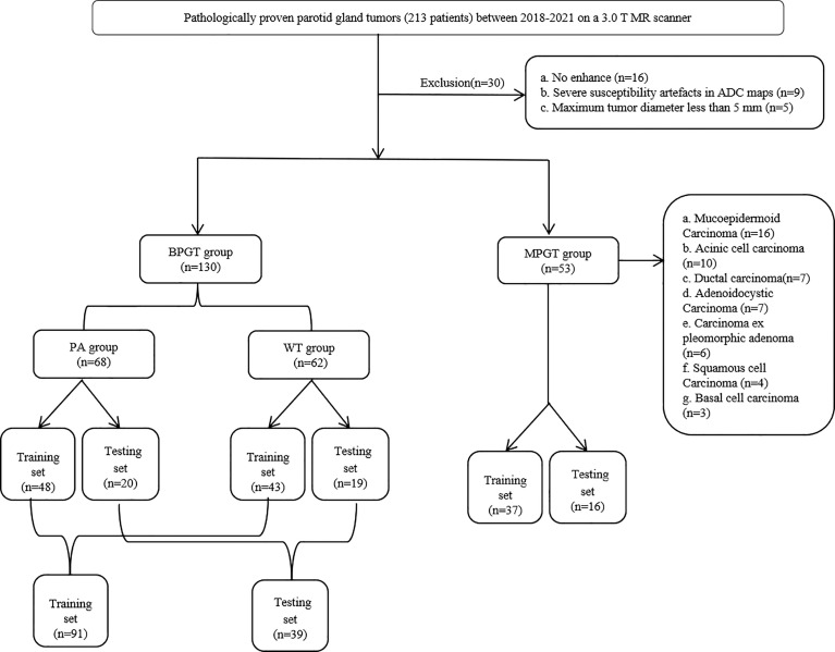
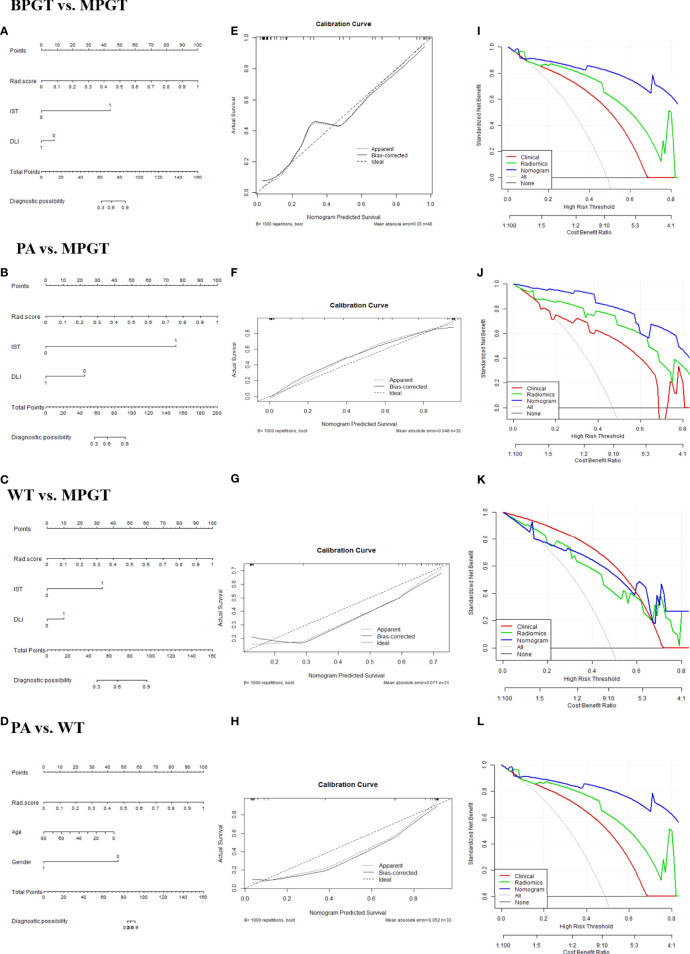
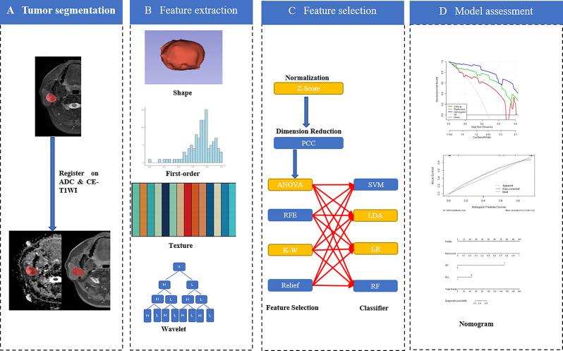
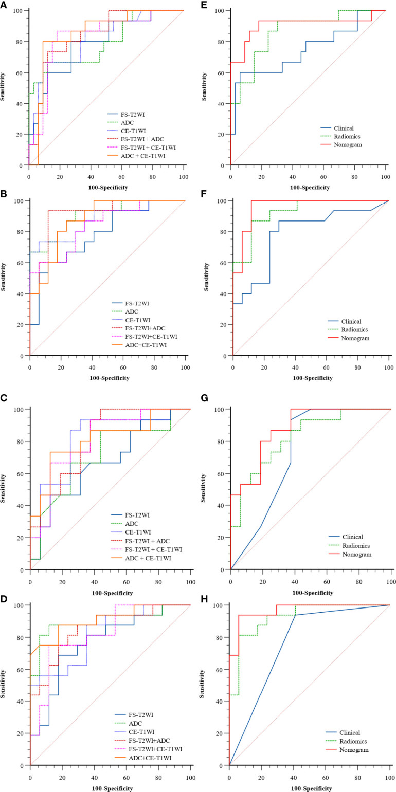
This retrospective study enrolled 183 parotid gland tumors (68 PAs, 62 WTs, and 53 MPGTs) and divided them into training (n = 128) and testing (n = 55) cohorts. In total, 2553 radiomics features were extracted from fat-saturated T2-weighted images, apparent diffusion coefficient maps, and contrast-enhanced T1-weighted images to construct single-, double-, and multi-sequence combined radiomics models, respectively. The radiomics score (Rad-score) was calculated using the best radiomics model and clinical features to develop the radiomics nomogram. The receiver operating characteristic curve and area under the curve (AUC) were used to assess these models, and their performances were compared using DeLong's test. Calibration curves and decision curve analysis were used to assess the clinical usefulness of these models.

RESULTS

The multi-sequence combined radiomics model exhibited better differentiation performance (BPGT . MPGT, AUC=0.863; PA . MPGT, AUC=0.929; WT . MPGT, AUC=0.825; PA . WT, AUC=0.927) than the single- and double sequence radiomics models. The nomogram based on the multi-sequence combined radiomics model and clinical features attained an improved classification performance (BPGT . MPGT, AUC=0.907; PA . MPGT, AUC=0.961; WT . MPGT, AUC=0.879; PA . WT, AUC=0.967).

CONCLUSIONS

Radiomics nomogram yielded excellent diagnostic performance in differentiating BPGT from MPGT, PA from MPGT, and PA from WT.

摘要

目的

我们旨在开发并验证放射组学列线图,以实现术前鉴别腮腺良性肿瘤和恶性肿瘤(分别为BPGT和MPGT),以及鉴别多形性腺瘤(PA)和沃辛瘤(WT)。

材料与方法

这项回顾性研究纳入了183例腮腺肿瘤(68例PA、62例WT和53例MPGT),并将它们分为训练队列(n = 128)和测试队列(n = 55)。总共从脂肪饱和T2加权图像、表观扩散系数图和对比增强T1加权图像中提取了2553个放射组学特征,分别构建单序列、双序列和多序列联合放射组学模型。使用最佳放射组学模型和临床特征计算放射组学评分(Rad-score),以建立放射组学列线图。采用受试者操作特征曲线和曲线下面积(AUC)评估这些模型,并使用德龙检验比较它们的性能。校准曲线和决策曲线分析用于评估这些模型的临床实用性。

结果

多序列联合放射组学模型表现出比单序列和双序列放射组学模型更好的鉴别性能(BPGT与MPGT相比,AUC = 0.863;PA与MPGT相比,AUC = 0.929;WT与MPGT相比,AUC = 0.825;PA与WT相比,AUC = 0.927)。基于多序列联合放射组学模型和临床特征的列线图获得了更好的分类性能(BPGT与MPGT相比,AUC = 0.907;PA与MPGT相比,AUC = 0.961;WT与MPGT相比,AUC = 0.879;PA与WT相比,AUC = 0.967)。

结论

放射组学列线图在鉴别BPGT与MPGT、PA与MPGT以及PA与WT方面具有出色的诊断性能。

https://cdn.ncbi.nlm.nih.gov/pmc/blobs/56b6/9309371/87c1a3bb0fd5/fonc-12-937050-g004.jpg
https://cdn.ncbi.nlm.nih.gov/pmc/blobs/56b6/9309371/92c2d90bbe32/fonc-12-937050-g001.jpg
https://cdn.ncbi.nlm.nih.gov/pmc/blobs/56b6/9309371/2eedd57dd8dc/fonc-12-937050-g002.jpg
https://cdn.ncbi.nlm.nih.gov/pmc/blobs/56b6/9309371/0d9902cf8ee6/fonc-12-937050-g003.jpg
https://cdn.ncbi.nlm.nih.gov/pmc/blobs/56b6/9309371/87c1a3bb0fd5/fonc-12-937050-g004.jpg

相似文献

1
Differentiation of Benign From Malignant Parotid Gland Tumors Using Conventional MRI Based on Radiomics Nomogram.基于影像组学列线图的传统MRI鉴别腮腺良恶性肿瘤
Front Oncol. 2022 Jul 11;12:937050. doi: 10.3389/fonc.2022.937050. eCollection 2022.
2
MRI-Based radiomics nomogram for differentiation of benign and malignant lesions of the parotid gland.基于 MRI 的影像组学列线图用于鉴别腮腺良恶性病变。
Eur Radiol. 2021 Jun;31(6):4042-4052. doi: 10.1007/s00330-020-07483-4. Epub 2020 Nov 19.
3
The Role of an MRI-Based Radiomic Signature in Predicting Malignancy of Parotid Gland Tumors.基于MRI的影像组学特征在预测腮腺肿瘤恶性程度中的作用
Cancers (Basel). 2023 Jun 23;15(13):3319. doi: 10.3390/cancers15133319.
4
Development and validation of CT-based radiomics nomogram for the classification of benign parotid gland tumors.基于 CT 的放射组学列线图构建与验证用于腮腺良恶性肿瘤分类
Med Phys. 2023 Feb;50(2):947-957. doi: 10.1002/mp.16042. Epub 2022 Nov 12.
5
CT-based radiomics with various classifiers for histological differentiation of parotid gland tumors.基于CT的放射组学结合多种分类器用于腮腺肿瘤的组织学鉴别
Front Oncol. 2023 Mar 10;13:1118351. doi: 10.3389/fonc.2023.1118351. eCollection 2023.
6
The value of T1- and FST2-Weighted-based radiomics nomogram in differentiating pleomorphic adenoma and Warthin tumor.基于T1加权和FST2加权的影像组学列线图在鉴别多形性腺瘤和沃辛瘤中的价值。
Transl Oncol. 2024 Nov;49:102087. doi: 10.1016/j.tranon.2024.102087. Epub 2024 Aug 18.
7
Distinguishing Parotid Polymorphic Adenoma and Warthin Tumor Based on the CT Radiomics Nomogram: A Multicenter Study.基于CT影像组学列线图鉴别腮腺多形性腺瘤和沃辛瘤:一项多中心研究
Acad Radiol. 2023 Apr;30(4):717-726. doi: 10.1016/j.acra.2022.06.017. Epub 2022 Aug 8.
8
Apparent Diffusion Coefficient Map-Based Radiomics Features for Differential Diagnosis of Pleomorphic Adenomas and Warthin Tumors From Malignant Tumors.基于表观扩散系数图的放射组学特征用于多形性腺瘤和沃辛瘤与恶性肿瘤的鉴别诊断
Front Oncol. 2022 Jun 7;12:830496. doi: 10.3389/fonc.2022.830496. eCollection 2022.
9
Enhanced CT-based texture analysis and radiomics score for differentiation of pleomorphic adenoma, basal cell adenoma, and Warthin tumor of the parotid gland.基于增强 CT 的纹理分析和放射组学评分在鉴别腮腺多形性腺瘤、基底细胞腺瘤和沃辛瘤中的应用。
Dentomaxillofac Radiol. 2023 Jan;52(2):20220009. doi: 10.1259/dmfr.20220009. Epub 2023 Jan 3.
10
Value of T2-weighted-based radiomics model in distinguishing Warthin tumor from pleomorphic adenoma of the parotid.基于 T2 加权的放射组学模型在鉴别腮腺沃辛瘤与多形性腺瘤中的价值。
Eur Radiol. 2023 Jun;33(6):4453-4463. doi: 10.1007/s00330-022-09295-0. Epub 2022 Dec 11.

引用本文的文献

1
Classification of Parotid Tumors with Robust Radiomic Features from DCE- and DW-MRI.利用动态对比增强磁共振成像(DCE-MRI)和扩散加权磁共振成像(DW-MRI)的稳健影像组学特征对腮腺肿瘤进行分类
J Imaging. 2025 Apr 17;11(4):122. doi: 10.3390/jimaging11040122.
2
MR Image Fusion-Based Parotid Gland Tumor Detection.基于磁共振图像融合的腮腺肿瘤检测
J Imaging Inform Med. 2025 Jun;38(3):1846-1859. doi: 10.1007/s10278-024-01137-3. Epub 2024 Sep 26.
3
Modified buried vertical mattress suture technique for excisional suturing of benign parotid tumors: A retrospective study.

本文引用的文献

1
Tumor blood flow and apparent diffusion coefficient histogram analysis for differentiating malignant salivary tumors from pleomorphic adenomas and Warthin's tumors.肿瘤血流和表观扩散系数直方图分析用于鉴别恶性唾液腺肿瘤、多形性腺瘤和沃辛瘤。
Sci Rep. 2022 Apr 8;12(1):5947. doi: 10.1038/s41598-022-09968-2.
2
Radiomics Analysis of Multiparametric MRI for Prediction of Synchronous Lung Metastases in Osteosarcoma.多参数MRI的影像组学分析用于预测骨肉瘤的同步肺转移
Front Oncol. 2022 Feb 22;12:802234. doi: 10.3389/fonc.2022.802234. eCollection 2022.
3
A nomogram based on clinical information, conventional ultrasound and radiomics improves prediction of malignant parotid gland lesions.
改良埋没垂直褥式缝合法用于腮腺良性肿瘤切除缝合的回顾性研究
J Cosmet Dermatol. 2024 Dec;23(12):4161-4166. doi: 10.1111/jocd.16521. Epub 2024 Aug 19.
4
The value of T1- and FST2-Weighted-based radiomics nomogram in differentiating pleomorphic adenoma and Warthin tumor.基于T1加权和FST2加权的影像组学列线图在鉴别多形性腺瘤和沃辛瘤中的价值。
Transl Oncol. 2024 Nov;49:102087. doi: 10.1016/j.tranon.2024.102087. Epub 2024 Aug 18.
5
Performance of radiomics in the differential diagnosis of parotid tumors: a systematic review.放射组学在腮腺肿瘤鉴别诊断中的应用:一项系统评价
Front Oncol. 2024 Jul 25;14:1383323. doi: 10.3389/fonc.2024.1383323. eCollection 2024.
6
Different MRI-based radiomics models for differentiating misdiagnosed or ambiguous pleomorphic adenoma and Warthin tumor of the parotid gland: a multicenter study.基于MRI的不同放射组学模型用于鉴别腮腺误诊或不明确的多形性腺瘤和沃辛瘤:一项多中心研究
Front Oncol. 2024 Jun 13;14:1392343. doi: 10.3389/fonc.2024.1392343. eCollection 2024.
7
Differentiation of benign and malignant parotid gland tumors based on the fusion of radiomics and deep learning features on ultrasound images.基于超声图像上的影像组学和深度学习特征融合对腮腺良恶性肿瘤进行鉴别诊断。
Front Oncol. 2024 May 13;14:1384105. doi: 10.3389/fonc.2024.1384105. eCollection 2024.
8
Radiomics Analysis in Characterization of Salivary Gland Tumors on MRI: A Systematic Review.MRI 对唾液腺肿瘤特征的影像组学分析:一项系统综述
Cancers (Basel). 2023 Oct 10;15(20):4918. doi: 10.3390/cancers15204918.
9
The Role of an MRI-Based Radiomic Signature in Predicting Malignancy of Parotid Gland Tumors.基于MRI的影像组学特征在预测腮腺肿瘤恶性程度中的作用
Cancers (Basel). 2023 Jun 23;15(13):3319. doi: 10.3390/cancers15133319.
10
CT-based radiomics with various classifiers for histological differentiation of parotid gland tumors.基于CT的放射组学结合多种分类器用于腮腺肿瘤的组织学鉴别
Front Oncol. 2023 Mar 10;13:1118351. doi: 10.3389/fonc.2023.1118351. eCollection 2023.
基于临床信息、常规超声和放射组学的列线图可提高腮腺良恶性病变的预测能力。
Cancer Lett. 2022 Feb 28;527:107-114. doi: 10.1016/j.canlet.2021.12.015. Epub 2021 Dec 17.
4
Radiomics for the Prediction of Epilepsy in Patients With Frontal Glioma.基于影像组学预测额叶胶质瘤患者的癫痫发作
Front Oncol. 2021 Nov 22;11:725926. doi: 10.3389/fonc.2021.725926. eCollection 2021.
5
Preoperative Magnetic Resonance Imaging Radiomics for Predicting Early Recurrence of Glioblastoma.术前磁共振成像放射组学预测胶质母细胞瘤早期复发
Front Oncol. 2021 Oct 27;11:769188. doi: 10.3389/fonc.2021.769188. eCollection 2021.
6
Applications of machine and deep learning to patient-specific IMRT/VMAT quality assurance.机器和深度学习在个体化调强放疗/容积旋转调强放疗质量保证中的应用。
J Appl Clin Med Phys. 2021 Sep;22(9):20-36. doi: 10.1002/acm2.13375. Epub 2021 Aug 3.
7
Differentiating low and high grade mucoepidermoid carcinoma of the salivary glands using CT radiomics.利用CT影像组学鉴别涎腺低级别和高级别黏液表皮样癌
Gland Surg. 2021 May;10(5):1646-1654. doi: 10.21037/gs-20-830.
8
MRI-Based Radiomics to Differentiate between Benign and Malignant Parotid Tumors With External Validation.基于磁共振成像的影像组学用于鉴别腮腺良恶性肿瘤并进行外部验证
Front Oncol. 2021 Apr 27;11:656918. doi: 10.3389/fonc.2021.656918. eCollection 2021.
9
Development and validation of an MRI-based radiomics nomogram for distinguishing Warthin's tumour from pleomorphic adenomas of the parotid gland.基于 MRI 的放射组学列线图开发和验证,用于鉴别腮腺沃辛瘤与多形性腺瘤。
Dentomaxillofac Radiol. 2021 Oct 1;50(7):20210023. doi: 10.1259/dmfr.20210023. Epub 2021 May 5.
10
Accuracy, predictability and prognostic implications of fine-needle aspiration biopsy for parotid gland tumours: A retrospective case series.细针抽吸活检对腮腺肿瘤的准确性、可预测性和预后意义:一项回顾性病例系列研究。
Clin Otolaryngol. 2021 Sep;46(5):1065-1072. doi: 10.1111/coa.13795. Epub 2021 May 31.