Institute for Biomagnetism and Biosignalanalysis, University of Münster, Münster, Germany.
Institute for Computational and Applied Mathematics, University of Münster, Münster, Germany.
Hum Brain Mapp. 2021 Mar;42(4):978-992. doi: 10.1002/hbm.25272. Epub 2020 Nov 6.
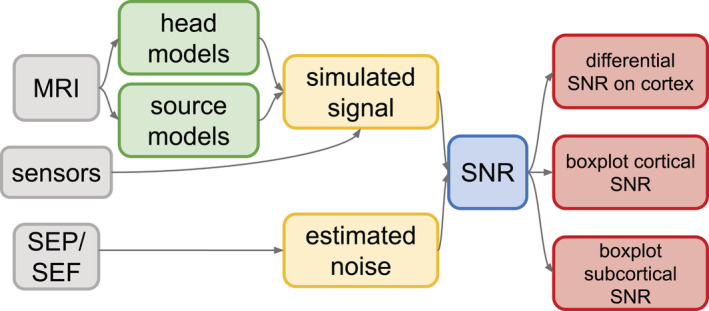
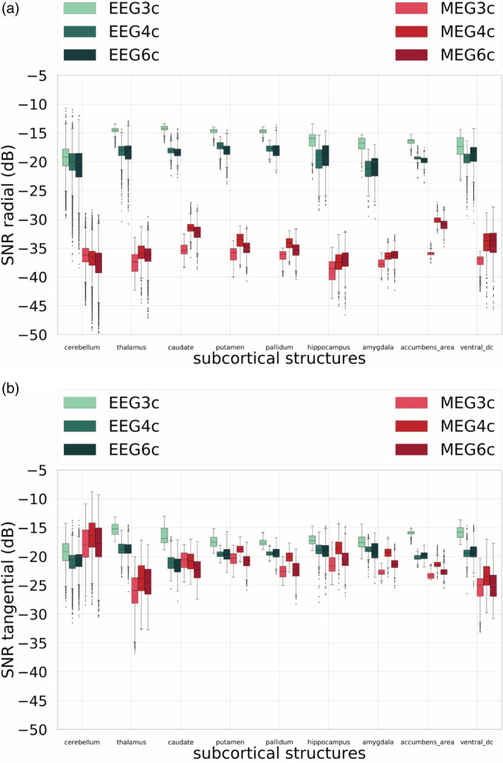
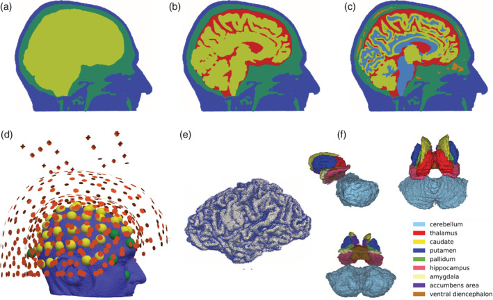
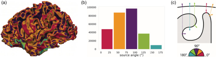
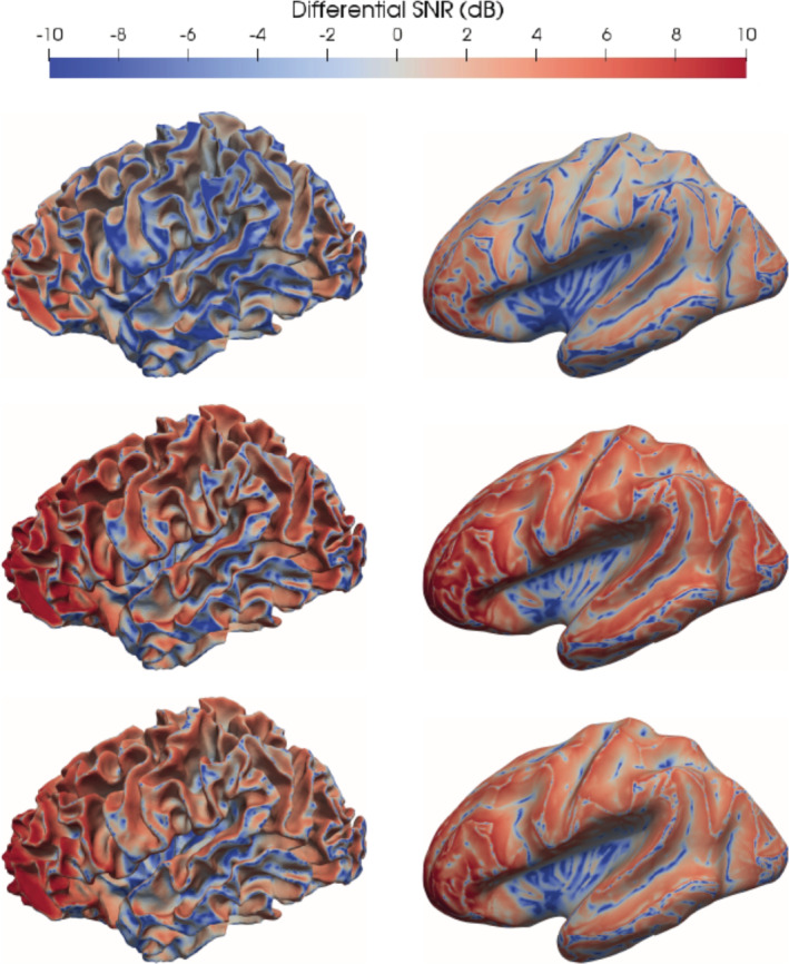
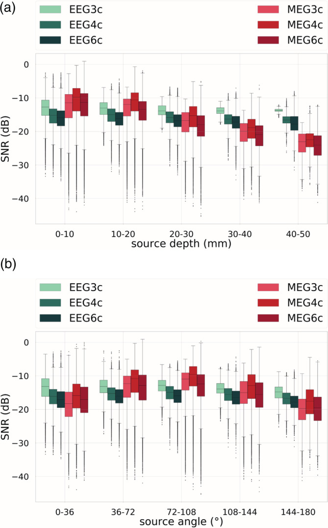
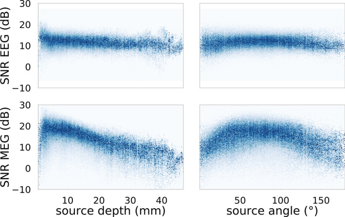
Signal-to-noise ratio (SNR) maps are a good way to visualize electroencephalography (EEG) and magnetoencephalography (MEG) sensitivity. SNR maps extend the knowledge about the modulation of EEG and MEG signals by source locations and orientations and can therefore help to better understand and interpret measured signals as well as source reconstruction results thereof. Our work has two main objectives. First, we investigated the accuracy and reliability of EEG and MEG finite element method (FEM)-based sensitivity maps for three different head models, namely an isotropic three and four-compartment and an anisotropic six-compartment head model. As a result, we found that ignoring the cerebrospinal fluid leads to an overestimation of EEG SNR values. Second, we examined and compared EEG and MEG SNR mappings for both cortical and subcortical sources and their modulation by source location and orientation. Our results for cortical sources show that EEG sensitivity is higher for radial and deep sources and MEG for tangential ones, which are the majority of sources. As to the subcortical sources, we found that deep sources with sufficient tangential source orientation are recordable by the MEG. Our work, which represents the first comprehensive study where cortical and subcortical sources are considered in highly detailed FEM-based EEG and MEG SNR mappings, sheds a new light on the sensitivity of EEG and MEG and might influence the decision of brain researchers or clinicians in their choice of the best modality for their experiment or diagnostics, respectively.
信噪比 (SNR) 图是可视化脑电图 (EEG) 和脑磁图 (MEG) 灵敏度的一种很好的方法。SNR 图扩展了关于 EEG 和 MEG 信号调制的知识,通过源位置和方向,因此可以帮助更好地理解和解释测量信号以及源重建结果。我们的工作有两个主要目标。首先,我们研究了三种不同头模型的基于 EEG 和 MEG 有限元方法 (FEM) 的灵敏度图的准确性和可靠性,即各向同性的三腔和四腔以及各向异性的六腔头模型。结果发现,忽略脑脊液会导致 EEG SNR 值的高估。其次,我们检查和比较了皮质和皮质下源的 EEG 和 MEG SNR 映射及其对源位置和方向的调制。我们对皮质源的结果表明,径向和深部源的 EEG 灵敏度较高,而切向源的 MEG 灵敏度较高,切向源是大多数源。至于皮质下源,我们发现具有足够切向源方向的深部源可以被 MEG 记录。我们的工作代表了第一个全面的研究,其中考虑了高度详细的基于 FEM 的 EEG 和 MEG SNR 映射中的皮质和皮质下源,为 EEG 和 MEG 的灵敏度提供了新的视角,并可能影响脑研究人员或临床医生在选择最适合其实验或诊断的最佳模态方面的决策。