Department of Companion and Laboratory Animal Science, Kongju National University, Yesan, Chungcheongnam 32439, Republic of Korea.
Department of Oral Pathology, School of Dentistry and Dental Research Institute, Seoul National University, Seoul 03080, Republic of Korea.
Mol Med Rep. 2020 Dec;22(6):4877-4889. doi: 10.3892/mmr.2020.11572. Epub 2020 Oct 8.
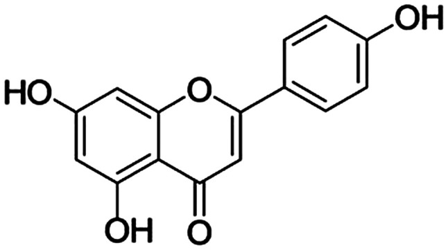
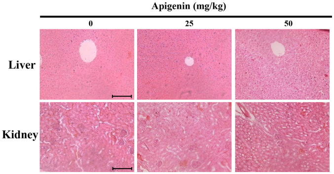
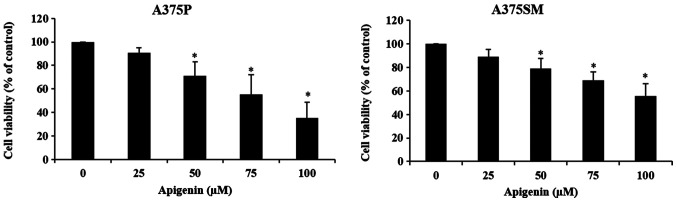
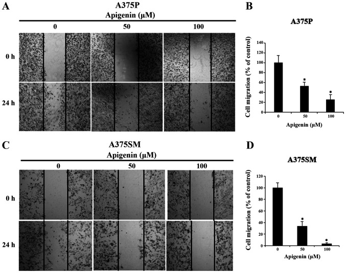
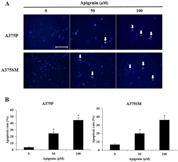
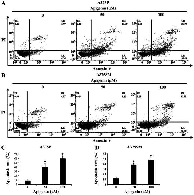
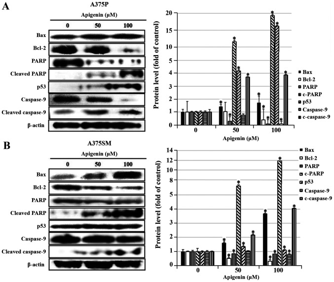
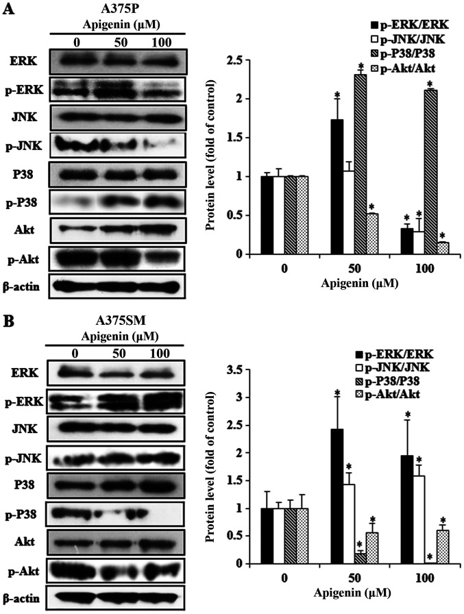
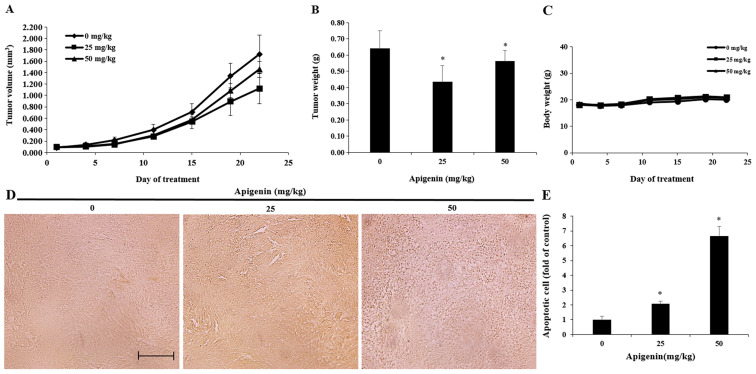
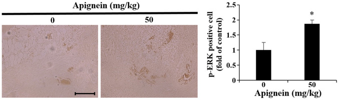
Apigenin, an aromatic compound, exhibits antioxidant, anti‑inflammatory and anti‑viral effects. The present study aimed to investigate the effects of apigenin on cell proliferation and apoptosis of human melanoma cells A375P and A375SM. Therefore, melanoma cells were treated with apigenin to determine its anti‑proliferative and survival effects, using wound healing and MTT assays. The results revealed that melanoma cell viability was decreased in a dose‑dependent manner. Furthermore, chromatin condensation, indicating apoptosis, was significantly increased in a dose‑dependent manner, as demonstrated by DAPI staining. In addition, increased apoptosis rate following treatment with apigenin was confirmed by Annexin V‑propidium iodide staining. The changes in the expression levels of apoptosis‑related proteins in A375P and A375SM melanoma cells were subsequently detected using western blot analysis. The results demonstrated that the protein expression levels of Bcl‑2 were decreased, whereas those of Bax, cleaved poly ADP‑ribose polymerase, cleaved caspase‑9 and p53 were upregulated in a dose‑dependent manner in apigenin‑treated cells compared with those noted in untreated cells. In addition, in apigenin‑treated A375P cells, phosphorylated (p)‑p38 was upregulated and p‑extracellular signal‑regulated kinase (ERK), p‑c‑Jun N‑terminal kinase (JNK) and p‑protein kinase B (Akt) were downregulated. However, in A375SM cells, apigenin treatment increased p‑ERK and p‑JNK and decreased p‑p38 and p‑Akt protein expression levels. Subsequently, the inhibitory effect of apigenin on tumor growth was investigated in vivo. Tumor volume was significantly reduced in the 25 and 50 mg/kg apigenin‑treated groups compared with the control group. Additionally, a TUNEL assay was performed to detect apoptotic cells. Immunohistochemical staining also revealed elevated p‑ERK expression in the apigenin‑treated group compared with the control group. Overall, the findings of the present study indicated that apigenin attenuated the growth of A375SM melanoma cells by inducing apoptosis via regulating the Akt and mitogen‑activated protein kinase signaling pathways.
芹菜素是一种芳香族化合物,具有抗氧化、抗炎和抗病毒作用。本研究旨在探讨芹菜素对人黑色素瘤细胞 A375P 和 A375SM 增殖和凋亡的影响。因此,用芹菜素处理黑色素瘤细胞,通过划痕愈合和 MTT 试验确定其抗增殖和生存作用。结果显示,黑色素瘤细胞活力呈剂量依赖性降低。此外,用 DAPI 染色显示,染色质浓缩,表明凋亡明显增加,呈剂量依赖性。此外,用 Annexin V-碘化丙啶染色证实,用芹菜素处理后凋亡率增加。随后通过 Western blot 分析检测 A375P 和 A375SM 黑色素瘤细胞中凋亡相关蛋白的表达水平变化。结果表明,与未处理细胞相比,芹菜素处理细胞中 Bcl-2 蛋白表达水平降低,而 Bax、裂解多聚 ADP-核糖聚合酶、裂解半胱天冬酶-9 和 p53 蛋白表达水平呈剂量依赖性增加。此外,在芹菜素处理的 A375P 细胞中,磷酸化 (p)-p38 上调,而 p-细胞外信号调节激酶 (ERK)、p-c-Jun N-末端激酶 (JNK) 和 p-蛋白激酶 B (Akt) 下调。然而,在 A375SM 细胞中,芹菜素处理增加了 p-ERK 和 p-JNK,降低了 p-p38 和 p-Akt 蛋白表达水平。随后,在体内研究了芹菜素对肿瘤生长的抑制作用。与对照组相比,25 和 50mg/kg 芹菜素处理组的肿瘤体积明显减小。此外,进行了 TUNEL 检测以检测凋亡细胞。免疫组化染色也显示,与对照组相比,芹菜素处理组 p-ERK 表达升高。总之,本研究结果表明,芹菜素通过调节 Akt 和丝裂原活化蛋白激酶信号通路诱导凋亡,从而减弱 A375SM 黑色素瘤细胞的生长。