Department of Biology, Technical University of Darmstadt, 64287 Darmstadt, Germany.
Department of Biology II, LMU Munich, 81377 Munich, Germany.
Nucleic Acids Res. 2020 Dec 16;48(22):12751-12777. doi: 10.1093/nar/gkaa1124.
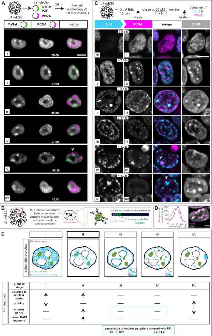
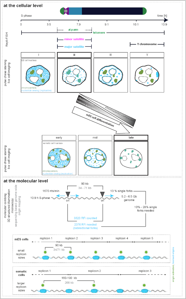
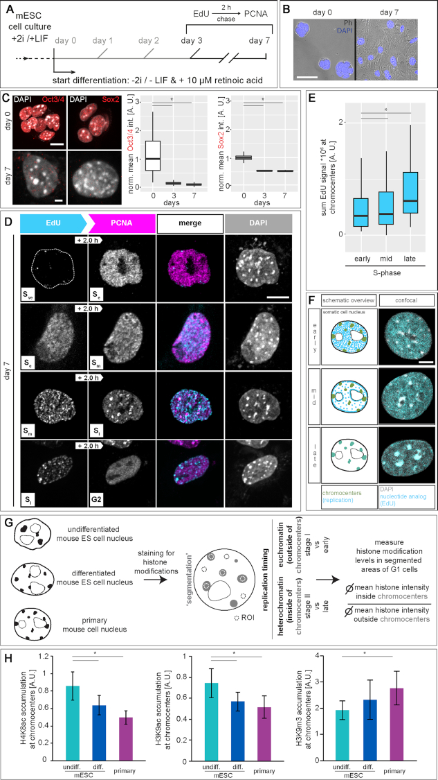
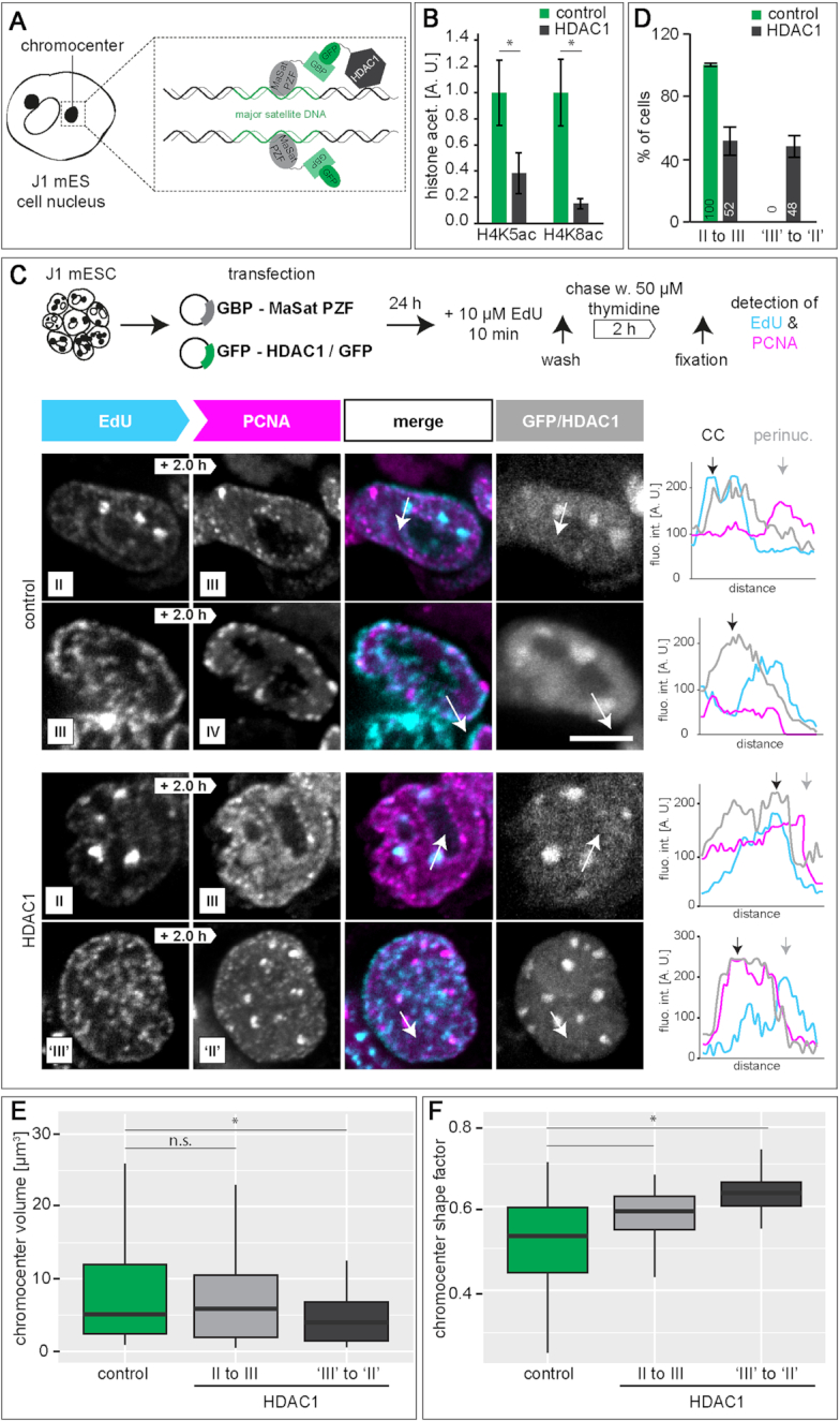
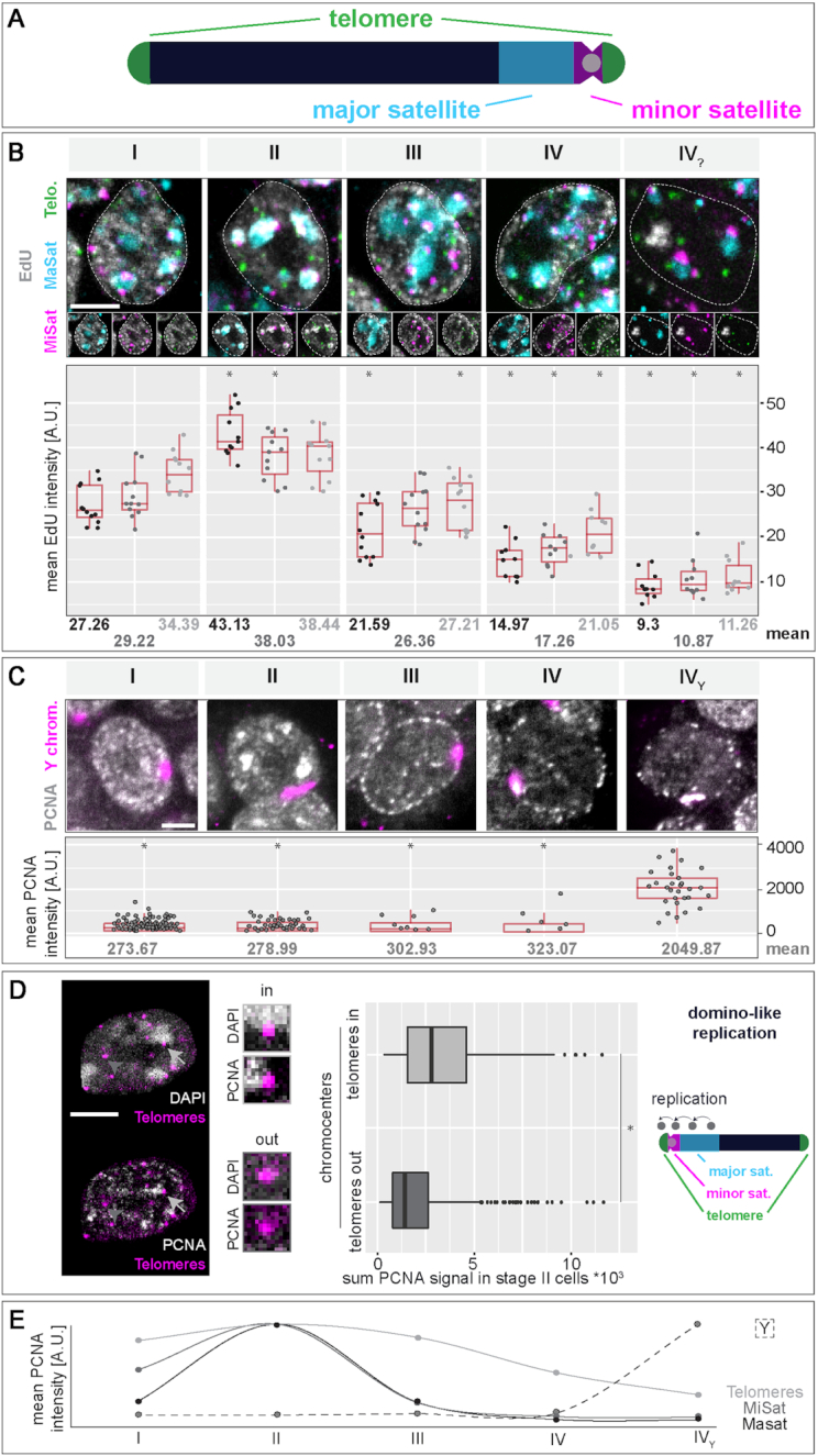
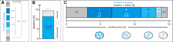
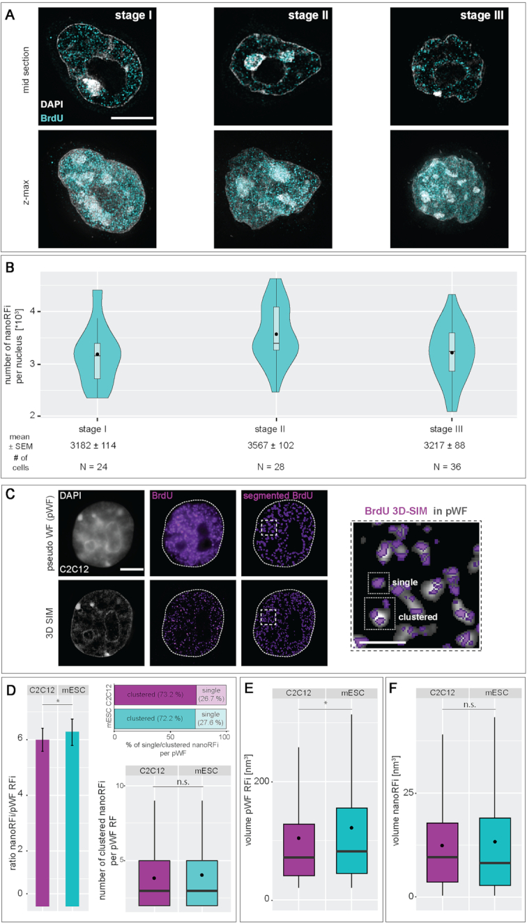
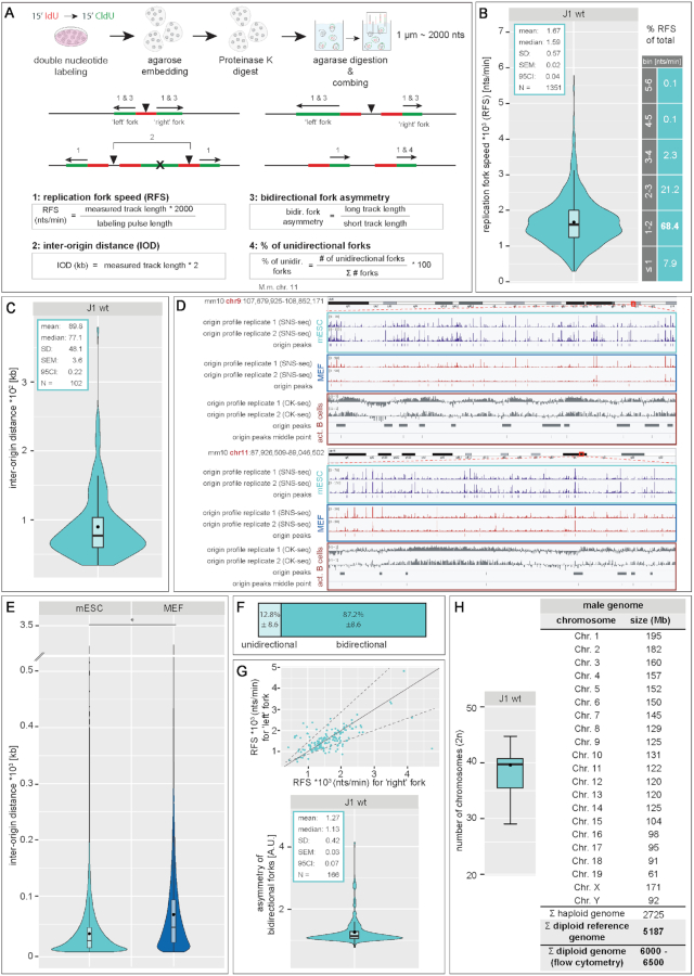
To ensure error-free duplication of all (epi)genetic information once per cell cycle, DNA replication follows a cell type and developmental stage specific spatio-temporal program. Here, we analyze the spatio-temporal DNA replication progression in (un)differentiated mouse embryonic stem (mES) cells. Whereas telomeres replicate throughout S-phase, we observe mid S-phase replication of (peri)centromeric heterochromatin in mES cells, which switches to late S-phase replication upon differentiation. This replication timing reversal correlates with and depends on an increase in condensation and a decrease in acetylation of chromatin. We further find synchronous duplication of the Y chromosome, marking the end of S-phase, irrespectively of the pluripotency state. Using a combination of single-molecule and super-resolution microscopy, we measure molecular properties of the mES cell replicon, the number of replication foci active in parallel and their spatial clustering. We conclude that each replication nanofocus in mES cells corresponds to an individual replicon, with up to one quarter representing unidirectional forks. Furthermore, with molecular combing and genome-wide origin mapping analyses, we find that mES cells activate twice as many origins spaced at half the distance than somatic cells. Altogether, our results highlight fundamental developmental differences on progression of genome replication and origin activation in pluripotent cells.
为了确保每个细胞周期内所有(表观)遗传信息的无差错复制,DNA 复制遵循细胞类型和发育阶段特异性的时空程序。在这里,我们分析了(未)分化的小鼠胚胎干细胞(mES)中 DNA 复制的时空进展。虽然端粒在整个 S 期都进行复制,但我们观察到 mES 细胞中(peri)着丝粒异染色质在 S 期中期进行复制,在分化后切换到晚期 S 期复制。这种复制时间的逆转与染色质的浓缩增加和乙酰化减少相关,并依赖于它们。我们进一步发现 Y 染色体的同步复制,标志着 S 期的结束,而与多能状态无关。我们使用单分子和超分辨率显微镜的组合,测量 mES 细胞复制子的分子特性、同时活跃的复制焦点的数量及其空间聚类。我们得出结论,mES 细胞中的每个复制纳米焦点对应于一个单独的复制子,其中多达四分之一代表单向叉。此外,通过分子梳理和全基因组起始点作图分析,我们发现 mES 细胞激活的起始点数量是体细胞的两倍,间隔是体细胞的一半。总之,我们的研究结果强调了多能细胞中基因组复制和起始点激活的进展方面存在的基本发育差异。