• 文献检索
  • 文档翻译
  • 深度研究
  • 学术资讯
  • Suppr Zotero 插件Zotero 插件
  • 邀请有礼
  • 套餐&价格
  • 历史记录
应用&插件
Suppr Zotero 插件Zotero 插件浏览器插件Mac 客户端Windows 客户端微信小程序
定价
高级版会员购买积分包购买API积分包
服务
文献检索文档翻译深度研究API 文档MCP 服务
关于我们
关于 Suppr公司介绍联系我们用户协议隐私条款
关注我们

Suppr 超能文献

核心技术专利:CN118964589B侵权必究
粤ICP备2023148730 号-1Suppr @ 2026

文献检索

告别复杂PubMed语法,用中文像聊天一样搜索,搜遍4000万医学文献。AI智能推荐,让科研检索更轻松。

立即免费搜索

文件翻译

保留排版,准确专业,支持PDF/Word/PPT等文件格式,支持 12+语言互译。

免费翻译文档

深度研究

AI帮你快速写综述,25分钟生成高质量综述,智能提取关键信息,辅助科研写作。

立即免费体验

利用基于深度学习的 4D 分割技术建立秀丽隐杆线虫胚胎形态图谱。

Establishment of a morphological atlas of the Caenorhabditis elegans embryo using deep-learning-based 4D segmentation.

机构信息

Department of Electrical Engineering, City University of Hong Kong, Hong Kong, 999077, China.

Center for Quantitative Biology, Peking University, 100871, Beijing, China.

出版信息

Nat Commun. 2020 Dec 7;11(1):6254. doi: 10.1038/s41467-020-19863-x.

DOI:10.1038/s41467-020-19863-x
PMID:33288755
原文链接:https://pmc.ncbi.nlm.nih.gov/articles/PMC7721714/
Abstract

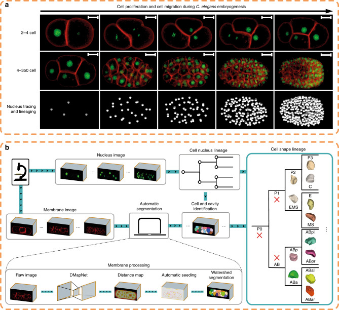
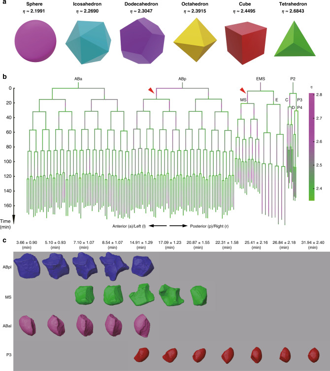
The invariant development and transparent body of the nematode Caenorhabditis elegans enables complete delineation of cell lineages throughout development. Despite extensive studies of cell division, cell migration and cell fate differentiation, cell morphology during development has not yet been systematically characterized in any metazoan, including C. elegans. This knowledge gap substantially hampers many studies in both developmental and cell biology. Here we report an automatic pipeline, CShaper, which combines automated segmentation of fluorescently labeled membranes with automated cell lineage tracing. We apply this pipeline to quantify morphological parameters of densely packed cells in 17 developing C. elegans embryos. Consequently, we generate a time-lapse 3D atlas of cell morphology for the C. elegans embryo from the 4- to 350-cell stages, including cell shape, volume, surface area, migration, nucleus position and cell-cell contact with resolved cell identities. We anticipate that CShaper and the morphological atlas will stimulate and enhance further studies in the fields of developmental biology, cell biology and biomechanics.

摘要

秀丽隐杆线虫(Caenorhabditis elegans)具有不变的发育和透明的身体,这使其能够在整个发育过程中完整描绘细胞谱系。尽管对细胞分裂、细胞迁移和细胞命运分化进行了广泛的研究,但在包括秀丽隐杆线虫在内的任何后生动物中,其发育过程中的细胞形态尚未得到系统描述。这一知识空白严重阻碍了发育生物学和细胞生物学领域的许多研究。在这里,我们报告了一个自动流水线 CShaper,它将荧光标记膜的自动分割与自动细胞谱系追踪相结合。我们将该流水线应用于在 17 个发育中的秀丽隐杆线虫胚胎中定量分析密集堆积细胞的形态参数。因此,我们生成了秀丽隐杆线虫胚胎从 4 细胞到 350 细胞阶段的延时 3D 细胞形态图谱,包括细胞形状、体积、表面积、迁移、核位置和细胞-细胞接触,同时解析了细胞身份。我们预计 CShaper 和形态图谱将激发和促进发育生物学、细胞生物学和生物力学领域的进一步研究。

https://cdn.ncbi.nlm.nih.gov/pmc/blobs/3484/7721714/f43281cc7d34/41467_2020_19863_Fig5_HTML.jpg
https://cdn.ncbi.nlm.nih.gov/pmc/blobs/3484/7721714/83d59b29e618/41467_2020_19863_Fig1_HTML.jpg
https://cdn.ncbi.nlm.nih.gov/pmc/blobs/3484/7721714/dd9d3c7aca1c/41467_2020_19863_Fig2_HTML.jpg
https://cdn.ncbi.nlm.nih.gov/pmc/blobs/3484/7721714/063294fc0643/41467_2020_19863_Fig3_HTML.jpg
https://cdn.ncbi.nlm.nih.gov/pmc/blobs/3484/7721714/38a99c721050/41467_2020_19863_Fig4_HTML.jpg
https://cdn.ncbi.nlm.nih.gov/pmc/blobs/3484/7721714/f43281cc7d34/41467_2020_19863_Fig5_HTML.jpg

相似文献

1
Establishment of a morphological atlas of the Caenorhabditis elegans embryo using deep-learning-based 4D segmentation.利用基于深度学习的 4D 分割技术建立秀丽隐杆线虫胚胎形态图谱。
Nat Commun. 2020 Dec 7;11(1):6254. doi: 10.1038/s41467-020-19863-x.
2
4D Microscopy: Unraveling Caenorhabditis elegans Embryonic Development Using Nomarski Microscopy.4D显微镜技术:利用诺马斯基显微镜解析秀丽隐杆线虫的胚胎发育
J Vis Exp. 2020 Oct 8(164). doi: 10.3791/61736.
3
Partially compromised specification causes stochastic effects on gut development in C. elegans.部分受损的规格对线虫肠道发育产生随机效应。
Dev Biol. 2017 Jul 1;427(1):49-60. doi: 10.1016/j.ydbio.2017.05.007. Epub 2017 May 11.
4
Timing of Tissue-specific Cell Division Requires a Differential Onset of Zygotic Transcription during Metazoan Embryogenesis.组织特异性细胞分裂的时间安排需要后生动物胚胎发生过程中合子转录的差异起始。
J Biol Chem. 2016 Jun 10;291(24):12501-12513. doi: 10.1074/jbc.M115.705426. Epub 2016 Apr 7.
5
Automated cell lineage reconstruction using label-free 4D microscopy.基于无标记 4D 显微镜的自动化细胞谱系重建。
Genetics. 2024 Oct 7;228(2). doi: 10.1093/genetics/iyae135.
6
The lineaging of fluorescently-labeled Caenorhabditis elegans embryos with StarryNite and AceTree.使用StarryNite和AceTree对荧光标记的秀丽隐杆线虫胚胎进行谱系追踪
Nat Protoc. 2006;1(3):1468-76. doi: 10.1038/nprot.2006.222.
7
Single-cell dynamics of chromatin activity during cell lineage differentiation in Caenorhabditis elegans embryos.秀丽隐杆线虫胚胎细胞谱系分化过程中染色质活性的单细胞动态变化。
Mol Syst Biol. 2021 Apr;17(4):e10075. doi: 10.15252/msb.202010075.
8
Cell lineage-resolved embryonic morphological map reveals signaling associated with cell fate and size asymmetry.细胞谱系解析的胚胎形态图谱揭示了与细胞命运和大小不对称相关的信号传导。
Nat Commun. 2025 Apr 18;16(1):3700. doi: 10.1038/s41467-025-58878-0.
9
Quantitative semi-automated analysis of morphogenesis with single-cell resolution in complex embryos.定量单细胞分辨率分析复杂胚胎中的形态发生。
Development. 2012 Nov;139(22):4271-9. doi: 10.1242/dev.086256. Epub 2012 Oct 10.
10
Multiple transcription factors directly regulate Hox gene lin-39 expression in ventral hypodermal cells of the C. elegans embryo and larva, including the hypodermal fate regulators LIN-26 and ELT-6.多种转录因子直接调控秀丽隐杆线虫胚胎和幼虫腹侧皮下细胞中的Hox基因lin-39表达,包括皮下命运调控因子LIN-26和ELT-6。
BMC Dev Biol. 2014 May 13;14:17. doi: 10.1186/1471-213X-14-17.

引用本文的文献

1
Cell lineage-resolved embryonic morphological map reveals signaling associated with cell fate and size asymmetry.细胞谱系解析的胚胎形态图谱揭示了与细胞命运和大小不对称相关的信号传导。
Nat Commun. 2025 Apr 18;16(1):3700. doi: 10.1038/s41467-025-58878-0.
2
Apico-basal intercalations enable the integrity of curved epithelia.顶端-基底间的嵌入连接确保了弯曲上皮组织的完整性。
Comput Struct Biotechnol J. 2025 Mar 19;27:1204-1214. doi: 10.1016/j.csbj.2025.03.011. eCollection 2025.
3
Deep learning-based aberration compensation improves contrast and resolution in fluorescence microscopy.

本文引用的文献

1
Cell lineage-dependent chiral actomyosin flows drive cellular rearrangements in early development.细胞谱系依赖性的手性肌动球蛋白流驱动早期发育中的细胞重排。
Elife. 2020 Jul 9;9:e54930. doi: 10.7554/eLife.54930.
2
A lineage-resolved molecular atlas of embryogenesis at single-cell resolution.单细胞分辨率解析胚胎发生的谱系分辨分子图谱。
Science. 2019 Sep 20;365(6459). doi: 10.1126/science.aax1971. Epub 2019 Sep 5.
3
Evaluation of Deep Learning Strategies for Nucleus Segmentation in Fluorescence Images.深度学习策略在荧光图像中细胞核分割的评估。
基于深度学习的像差补偿提高了荧光显微镜的对比度和分辨率。
Nat Commun. 2025 Jan 2;16(1):313. doi: 10.1038/s41467-024-55267-x.
4
Deep learning-based enhancement of fluorescence labeling for accurate cell lineage tracing during embryogenesis.基于深度学习的荧光标记增强技术在胚胎发生过程中进行精确的细胞谱系追踪。
Bioinformatics. 2024 Nov 1;40(11). doi: 10.1093/bioinformatics/btae626.
5
Computational Methods for Image Analysis in Craniofacial Development and Disease.颅面发育与疾病中图像分析的计算方法
J Dent Res. 2024 Dec;103(13):1340-1348. doi: 10.1177/00220345241265048. Epub 2024 Sep 13.
6
Automated cell lineage reconstruction using label-free 4D microscopy.基于无标记 4D 显微镜的自动化细胞谱系重建。
Genetics. 2024 Oct 7;228(2). doi: 10.1093/genetics/iyae135.
7
Advancing insights into microgravity induced muscle changes using Caenorhabditis elegans as a model organism.以秀丽隐杆线虫作为模式生物,深入了解微重力引起的肌肉变化。
NPJ Microgravity. 2024 Jul 26;10(1):79. doi: 10.1038/s41526-024-00418-z.
8
Characterizing Cellular Physiological States with Three-Dimensional Shape Descriptors for Cell Membranes.利用细胞膜的三维形状描述符表征细胞生理状态
Membranes (Basel). 2024 Jun 7;14(6):137. doi: 10.3390/membranes14060137.
9
A mathematical framework for understanding the spontaneous emergence of complexity applicable to growing multicellular systems.用于理解适用于不断增长的多细胞系统的复杂性自发出现的数学框架。
PLoS Comput Biol. 2024 Jun 5;20(6):e1011882. doi: 10.1371/journal.pcbi.1011882. eCollection 2024 Jun.
10
Universal consensus 3D segmentation of cells from 2D segmented stacks.从二维分割堆栈中对细胞进行通用一致的三维分割。
bioRxiv. 2025 Mar 20:2024.05.03.592249. doi: 10.1101/2024.05.03.592249.
Cytometry A. 2019 Sep;95(9):952-965. doi: 10.1002/cyto.a.23863. Epub 2019 Jul 16.
4
Applications, promises, and pitfalls of deep learning for fluorescence image reconstruction.深度学习在荧光图像重建中的应用、前景与挑战。
Nat Methods. 2019 Dec;16(12):1215-1225. doi: 10.1038/s41592-019-0458-z. Epub 2019 Jul 8.
5
MorphoNet: an interactive online morphological browser to explore complex multi-scale data.MorphoNet:一个交互式在线形态学浏览器,用于探索复杂的多尺度数据。
Nat Commun. 2019 Jun 27;10(1):2812. doi: 10.1038/s41467-019-10668-1.
6
Deep learning for cellular image analysis.深度学习在细胞图像分析中的应用。
Nat Methods. 2019 Dec;16(12):1233-1246. doi: 10.1038/s41592-019-0403-1. Epub 2019 May 27.
7
Specific Interactions Between Autosome and Chromosomes Cause Hybrid Male Sterility in Species.常染色体与染色体的特异性相互作用导致物种杂种雄性不育。
Genetics. 2019 Jul;212(3):801-813. doi: 10.1534/genetics.119.302202. Epub 2019 May 7.
8
Learn to segment single cells with deep distance estimator and deep cell detector.学习使用深度距离估计器和深度细胞探测器对单细胞进行分割。
Comput Biol Med. 2019 May;108:133-141. doi: 10.1016/j.compbiomed.2019.04.006. Epub 2019 Apr 8.
9
3DMMS: robust 3D Membrane Morphological Segmentation of C. elegans embryo.3DMMS:秀丽隐杆线虫胚胎的稳健三维细胞膜形态分割。
BMC Bioinformatics. 2019 Apr 8;20(1):176. doi: 10.1186/s12859-019-2720-x.
10
High-throughput, high-resolution deep learning microscopy based on registration-free generative adversarial network.基于无配准生成对抗网络的高通量、高分辨率深度学习显微镜技术。
Biomed Opt Express. 2019 Feb 4;10(3):1044-1063. doi: 10.1364/BOE.10.001044. eCollection 2019 Mar 1.