Institute for Medical Engineering and Science, MIT, Cambridge, MA, USA.
Picower Institute for Learning and Memory, MIT, Cambridge, MA, USA.
Sci Rep. 2020 Dec 8;10(1):21487. doi: 10.1038/s41598-020-78130-7.
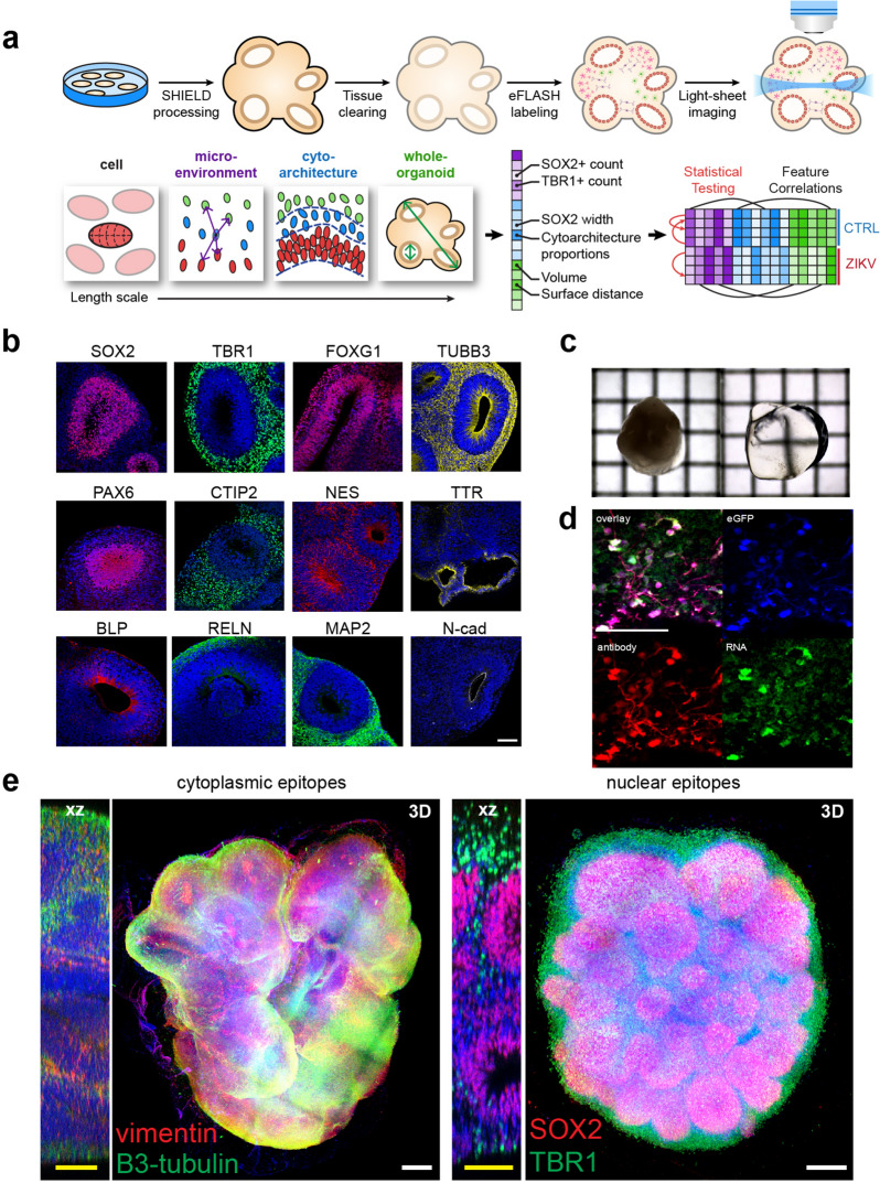
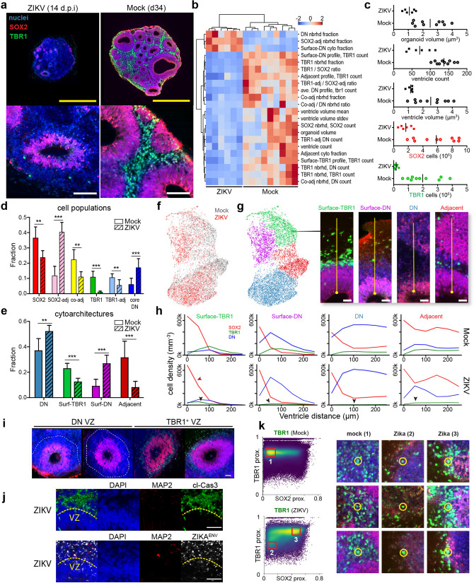
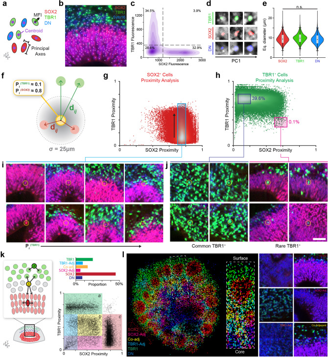
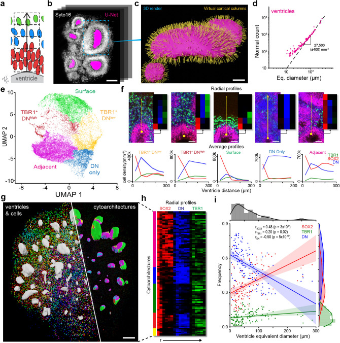
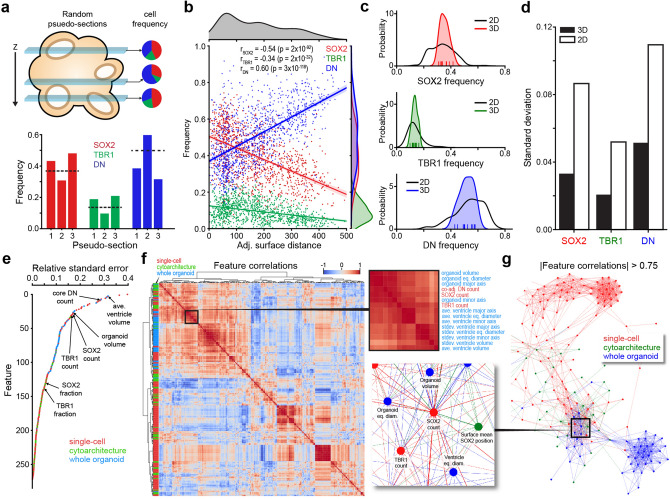
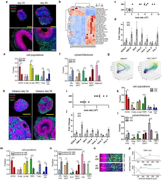
Brain organoids grown from human pluripotent stem cells self-organize into cytoarchitectures resembling the developing human brain. These three-dimensional models offer an unprecedented opportunity to study human brain development and dysfunction. Characterization currently sacrifices spatial information for single-cell or histological analysis leaving whole-tissue analysis mostly unexplored. Here, we present the SCOUT pipeline for automated multiscale comparative analysis of intact cerebral organoids. Our integrated technology platform can rapidly clear, label, and image intact organoids. Algorithmic- and convolutional neural network-based image analysis extract hundreds of features characterizing molecular, cellular, spatial, cytoarchitectural, and organoid-wide properties from fluorescence microscopy datasets. Comprehensive analysis of 46 intact organoids and ~ 100 million cells reveals quantitative multiscale "phenotypes" for organoid development, culture protocols and Zika virus infection. SCOUT provides a much-needed framework for comparative analysis of emerging 3D in vitro models using fluorescence microscopy.
由人类多能干细胞生长而成的大脑类器官会自行组织成类似于人类大脑发育过程的细胞结构。这些三维模型为研究人类大脑发育和功能障碍提供了前所未有的机会。目前的特征描述方法为了进行单细胞或组织学分析而牺牲了空间信息,使得对整个组织的分析大多仍未被探索。在这里,我们提出了 SCOUT 流水线,用于对完整的大脑类器官进行自动化多尺度比较分析。我们的集成技术平台可以快速对完整的类器官进行清洗、标记和成像。基于算法和卷积神经网络的图像分析从荧光显微镜数据集中提取数百个特征,这些特征可用于描述分子、细胞、空间、细胞结构和类器官整体特性。对 46 个完整的类器官和大约 1 亿个细胞进行的综合分析揭示了类器官发育、培养方案和寨卡病毒感染的定量多尺度“表型”。SCOUT 为使用荧光显微镜对新兴的 3D 体外模型进行比较分析提供了急需的框架。