Department of Biological Engineering, MIT, Cambridge, MA, USA.
Stanley Center for Psychiatric Research, Broad Institute of MIT and Harvard, Cambridge, MA, USA.
Nat Commun. 2019 Sep 26;10(1):4377. doi: 10.1038/s41467-019-12372-6.
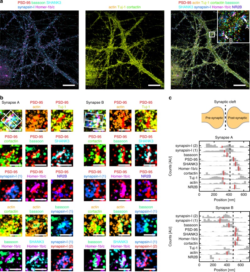
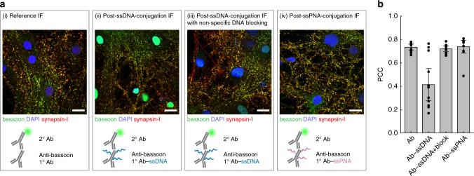
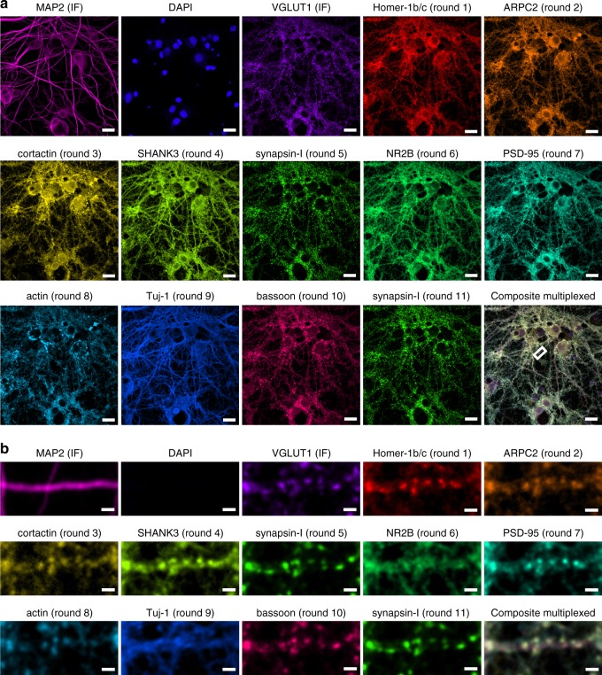
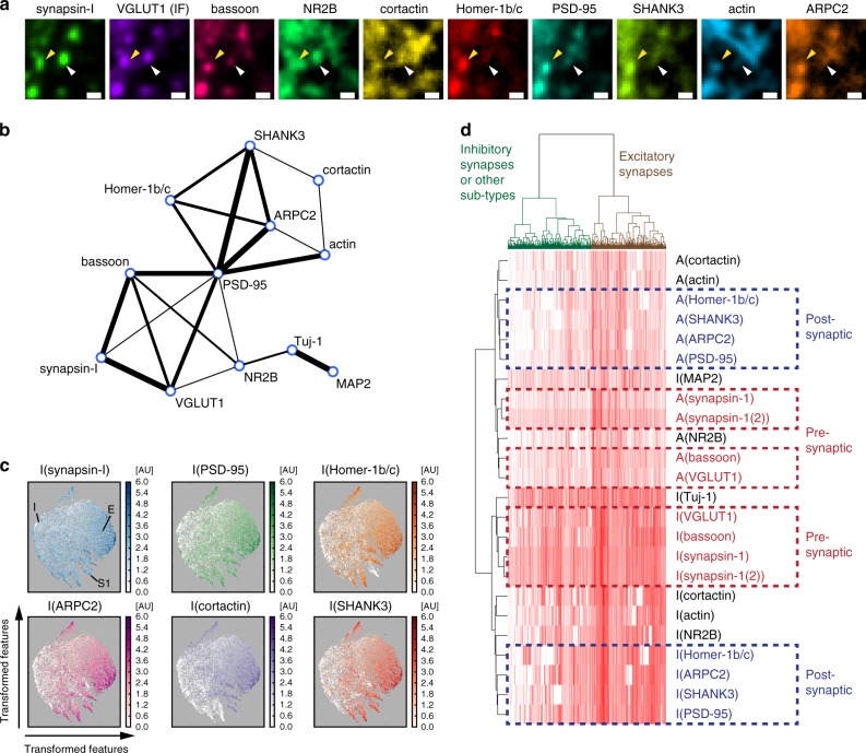
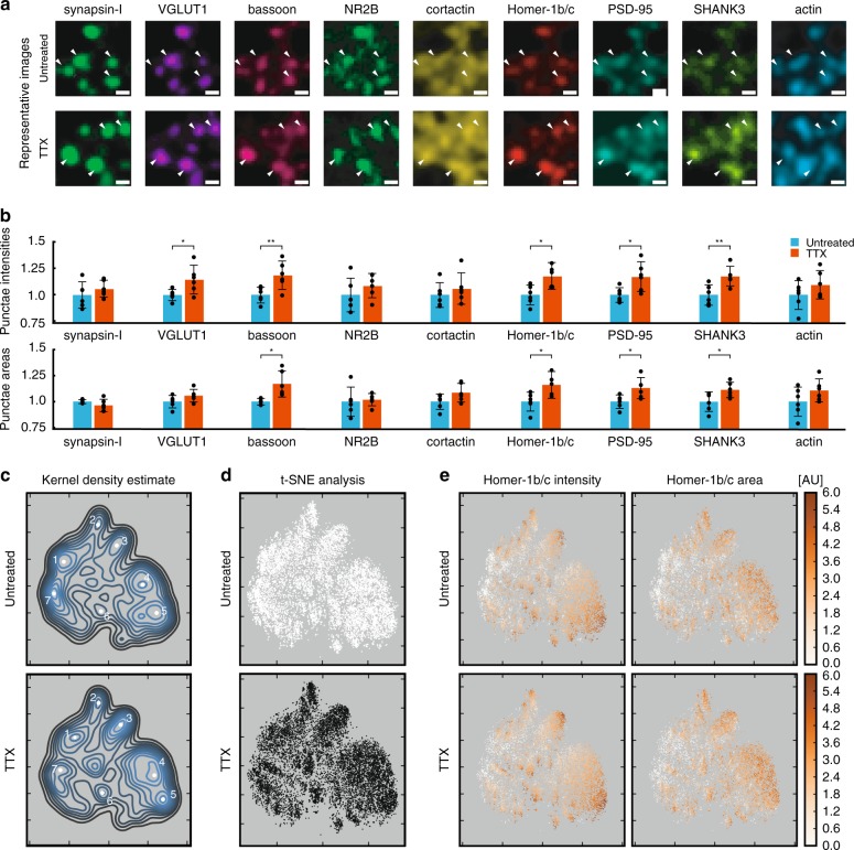
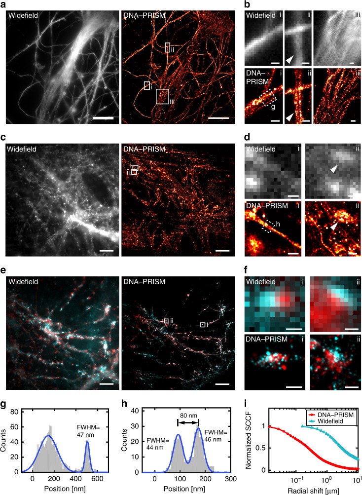
Synapses contain hundreds of distinct proteins whose heterogeneous expression levels are determinants of synaptic plasticity and signal transmission relevant to a range of diseases. Here, we use diffusible nucleic acid imaging probes to profile neuronal synapses using multiplexed confocal and super-resolution microscopy. Confocal imaging is performed using high-affinity locked nucleic acid imaging probes that stably yet reversibly bind to oligonucleotides conjugated to antibodies and peptides. Super-resolution PAINT imaging of the same targets is performed using low-affinity DNA imaging probes to resolve nanometer-scale synaptic protein organization across nine distinct protein targets. Our approach enables the quantitative analysis of thousands of synapses in neuronal culture to identify putative synaptic sub-types and co-localization patterns from one dozen proteins. Application to characterize synaptic reorganization following neuronal activity blockade reveals coordinated upregulation of the post-synaptic proteins PSD-95, SHANK3 and Homer-1b/c, as well as increased correlation between synaptic markers in the active and synaptic vesicle zones.
突触包含数百种不同的蛋白质,其异质表达水平是决定突触可塑性和信号传递的因素,与一系列疾病有关。在这里,我们使用可扩散核酸成像探针,通过共聚焦和超分辨率显微镜对神经元突触进行多指标分析。共聚焦成像使用高亲和力的锁核酸成像探针进行,该探针稳定但可逆地结合到与抗体和肽偶联的寡核苷酸上。使用低亲和力 DNA 成像探针对同一靶标进行超分辨率 PAINT 成像,以解析跨越九个不同蛋白质靶标的纳米级突触蛋白组织。我们的方法能够对神经元培养物中的数千个突触进行定量分析,以识别数十种蛋白质的潜在突触亚型和共定位模式。应用于描述神经元活动阻断后的突触重组,揭示了突触后蛋白 PSD-95、SHANK3 和 Homer-1b/c 的协调上调,以及活性区和突触小泡区的突触标记物之间相关性的增加。