MRILab, Institute for Molecular Imaging and Instrumentation (i3M), Spanish National Research Council (CSIC) and Universitat Politècnica de València (UPV), 46022, Valencia, Spain.
Tesoro Imaging S.L., 46022, Valencia, Spain.
Sci Rep. 2020 Dec 8;10(1):21470. doi: 10.1038/s41598-020-78456-2.
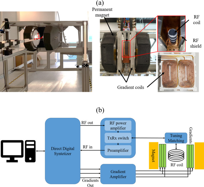
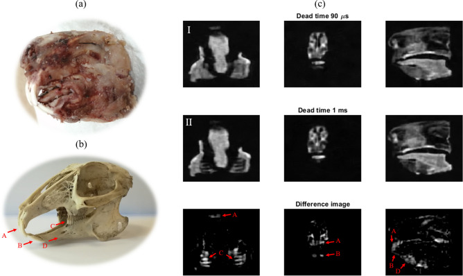
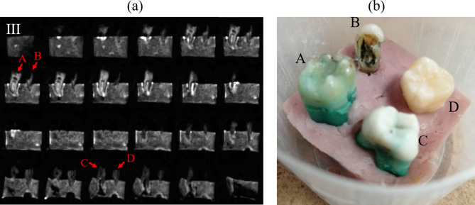
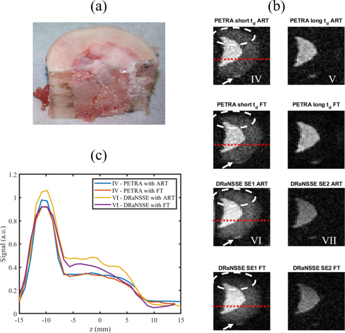
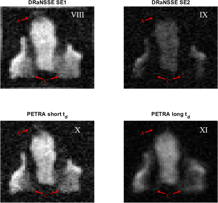
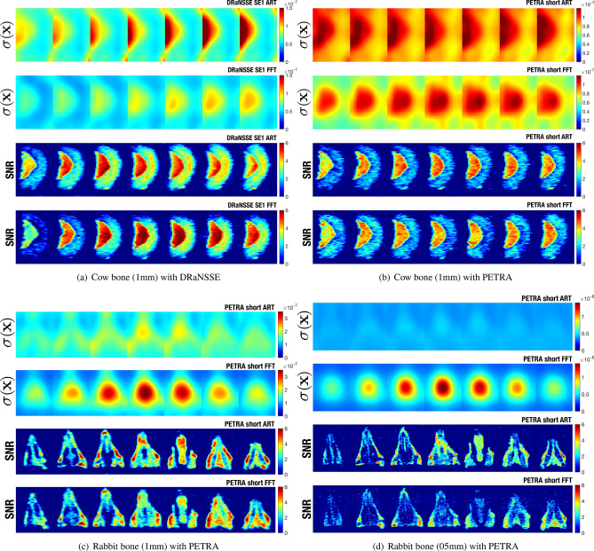
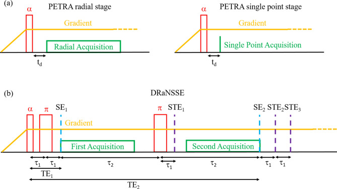
Magnetic Resonance Imaging (MRI) of hard biological tissues is challenging due to the fleeting lifetime and low strength of their response to resonant stimuli, especially at low magnetic fields. Consequently, the impact of MRI on some medical applications, such as dentistry, continues to be limited. Here, we present three-dimensional reconstructions of ex-vivo human teeth, as well as a rabbit head and part of a cow femur, all obtained at a field strength of 260 mT. These images are the first featuring soft and hard tissues simultaneously at sub-Tesla fields, and they have been acquired in a home-made, special-purpose, pre-medical MRI scanner designed with the goal of demonstrating dental imaging at low field settings. We encode spatial information with two pulse sequences: Pointwise-Encoding Time reduction with Radial Acquisition and a new sequence we have called Double Radial Non-Stop Spin Echo, which we find to perform better than the former. For image reconstruction we employ Algebraic Reconstruction Techniques (ART) as well as standard Fourier methods. An analysis of the resulting images shows that ART reconstructions exhibit a higher signal-to-noise ratio with a more homogeneous noise distribution.
硬生物组织的磁共振成像(MRI)具有挑战性,因为它们对共振刺激的响应寿命短暂且强度低,尤其是在低磁场下。因此,MRI 在一些医学应用中的影响,如牙科,仍然受到限制。在这里,我们展示了在 260 mT 场强下获得的离体人牙、兔头和部分牛股骨的三维重建。这些图像是首次在亚特斯拉场同时显示软、硬组织的图像,它们是在我们自行设计的、专用的、预医疗 MRI 扫描仪中获得的,旨在展示低场设置下的牙科成像。我们使用两种脉冲序列来编码空间信息:点编码时间减少的径向采集和我们称为双径向不停自旋回波的新序列,我们发现后者比前者表现更好。对于图像重建,我们采用代数重建技术(ART)和标准傅里叶方法。对所得图像的分析表明,ART 重建具有更高的信噪比和更均匀的噪声分布。