Saunders Diane C, Messmer James, Kusmartseva Irina, Beery Maria L, Yang Mingder, Atkinson Mark A, Powers Alvin C, Cartailler Jean-Philippe, Brissova Marcela
Division of Diabetes, Endocrinology, and Metabolism, Department of Medicine, Vanderbilt University Medical Center, Nashville, TN, USA.
Department of Pathology, Immunology, and Laboratory Medicine, College of Medicine, Diabetes Institute, University of Florida, Gainesville, FL, USA.
Patterns (N Y). 2020 Oct 5;1(8):100120. doi: 10.1016/j.patter.2020.100120. eCollection 2020 Nov 13.
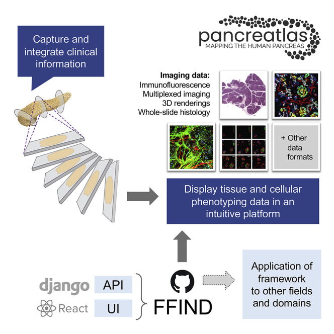
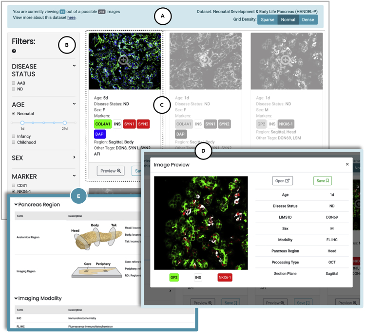
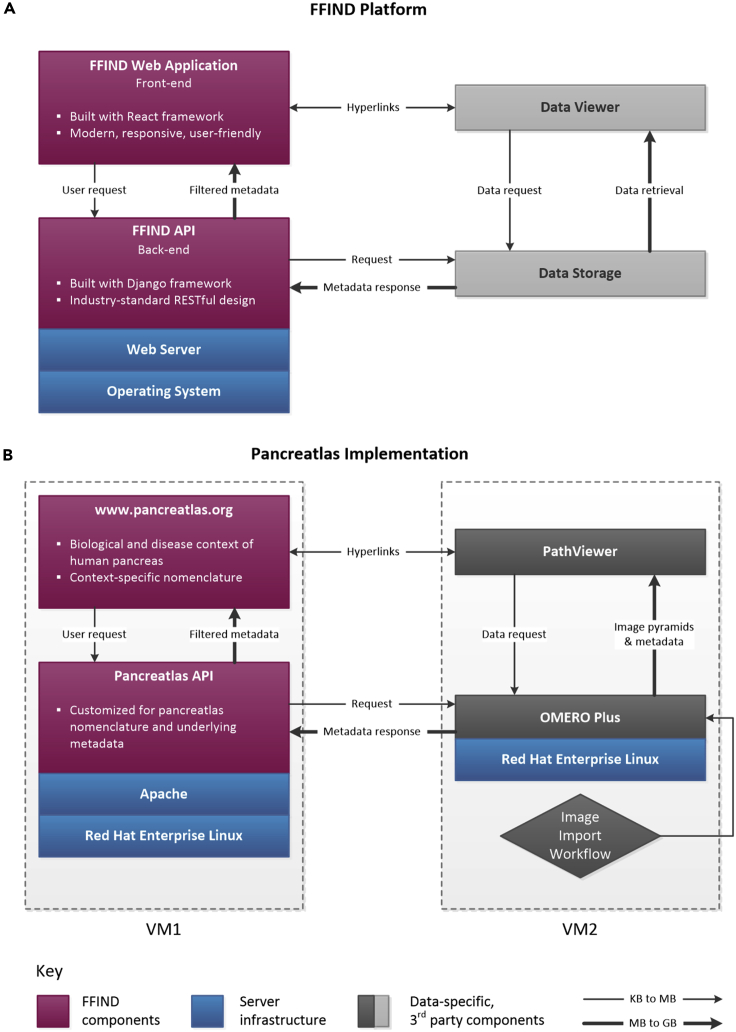
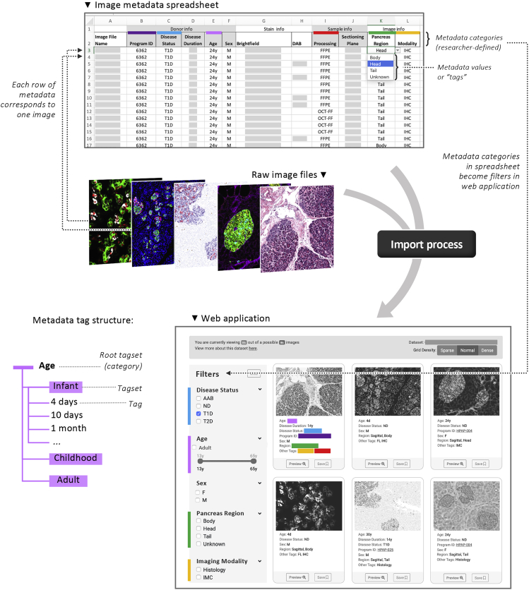
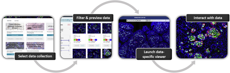
Human tissue phenotyping generates complex spatial information from numerous imaging modalities, yet images typically become static figures for publication, and original data and metadata are rarely available. While comprehensive image maps exist for some organs, most resources have limited support for multiplexed imaging or have non-intuitive user interfaces. Therefore, we built a Pancreatlas resource that integrates several technologies into a unique interface, allowing users to access richly annotated web pages, drill down to individual images, and deeply explore data online. The current version of Pancreatlas contains over 800 unique images acquired by whole-slide scanning, confocal microscopy, and imaging mass cytometry, and is available at https://www.pancreatlas.org. To create this human pancreas-specific biological imaging resource, we developed a React-based web application and Python-based application programming interface, collectively called Flexible Framework for Integrating and Navigating Data (FFIND), which can be adapted beyond Pancreatlas to meet countless imaging or other structured data-management needs.
人体组织表型分析可从众多成像方式中生成复杂的空间信息,但图像通常会成为用于发表的静态图,原始数据和元数据很少能获取到。虽然某些器官有全面的图像图谱,但大多数资源对多重成像的支持有限,或者用户界面不直观。因此,我们构建了一个胰腺图谱资源,将多种技术集成到一个独特的界面中,允许用户访问注释丰富的网页,深入查看单个图像,并在线深入探索数据。胰腺图谱的当前版本包含通过全切片扫描、共聚焦显微镜和成像质谱流式细胞术获取的800多张独特图像,可在https://www.pancreatlas.org上获取。为了创建这个特定于人类胰腺的生物成像资源,我们开发了一个基于React的Web应用程序和基于Python的应用程序编程接口,统称为数据集成与导航灵活框架(FFIND),它可以超越胰腺图谱进行调整,以满足无数成像或其他结构化数据管理需求。