• 文献检索
  • 文档翻译
  • 深度研究
  • 学术资讯
  • Suppr Zotero 插件Zotero 插件
  • 邀请有礼
  • 套餐&价格
  • 历史记录
应用&插件
Suppr Zotero 插件Zotero 插件浏览器插件Mac 客户端Windows 客户端微信小程序
定价
高级版会员购买积分包购买API积分包
服务
文献检索文档翻译深度研究API 文档MCP 服务
关于我们
关于 Suppr公司介绍联系我们用户协议隐私条款
关注我们

Suppr 超能文献

核心技术专利:CN118964589B侵权必究
粤ICP备2023148730 号-1Suppr @ 2026

文献检索

告别复杂PubMed语法,用中文像聊天一样搜索,搜遍4000万医学文献。AI智能推荐,让科研检索更轻松。

立即免费搜索

文件翻译

保留排版,准确专业,支持PDF/Word/PPT等文件格式,支持 12+语言互译。

免费翻译文档

深度研究

AI帮你快速写综述,25分钟生成高质量综述,智能提取关键信息,辅助科研写作。

立即免费体验

4Pi-SMS 超分辨率显微镜的实现。

Implementation of a 4Pi-SMS super-resolution microscope.

机构信息

Department of Engineering Science, University of Oxford, Oxford, UK.

The Gurdon Institute, University of Cambridge, Cambridge, UK.

出版信息

Nat Protoc. 2021 Feb;16(2):677-727. doi: 10.1038/s41596-020-00428-7. Epub 2020 Dec 16.

DOI:10.1038/s41596-020-00428-7
PMID:33328610
原文链接:https://pmc.ncbi.nlm.nih.gov/articles/PMC9118368/
Abstract

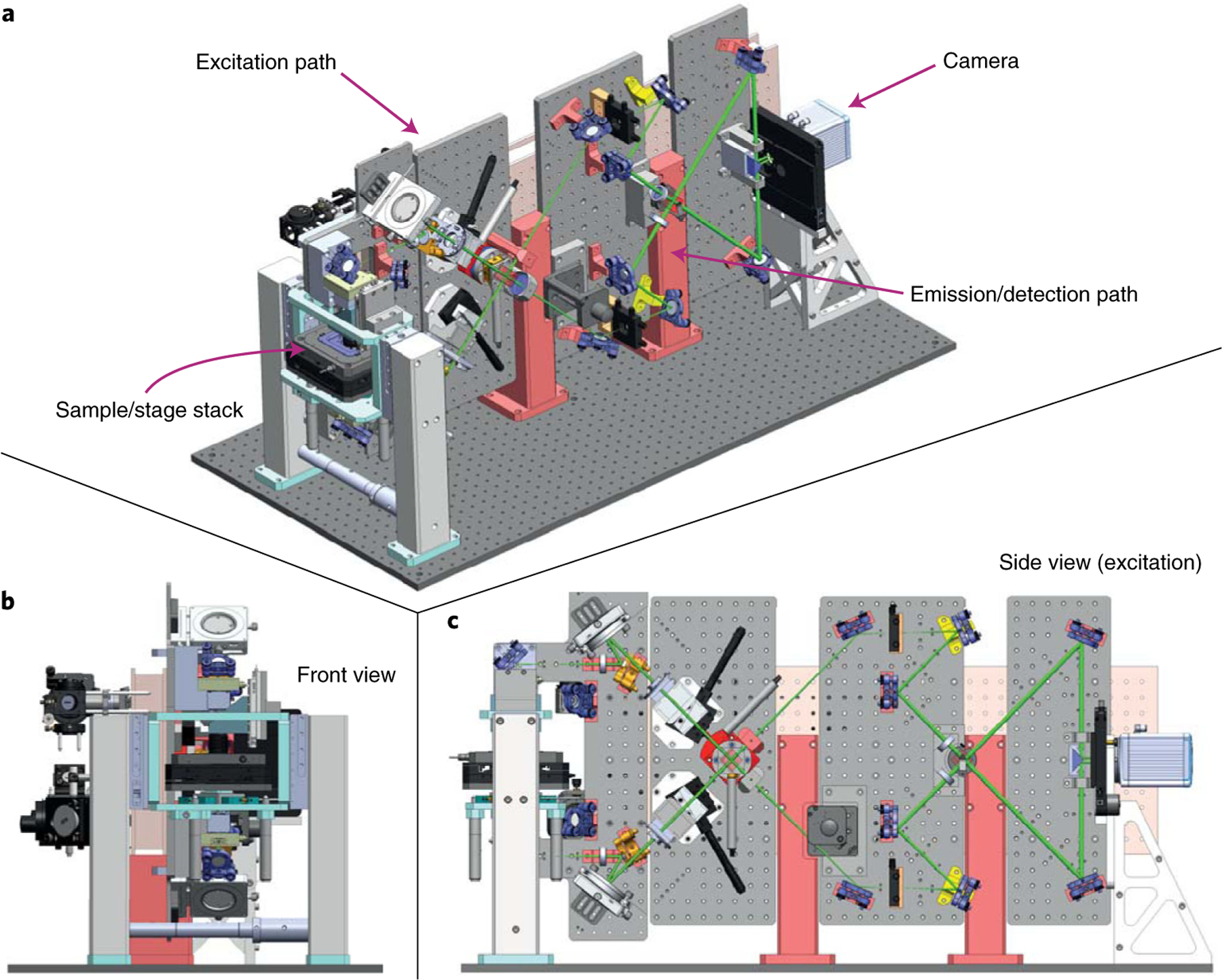
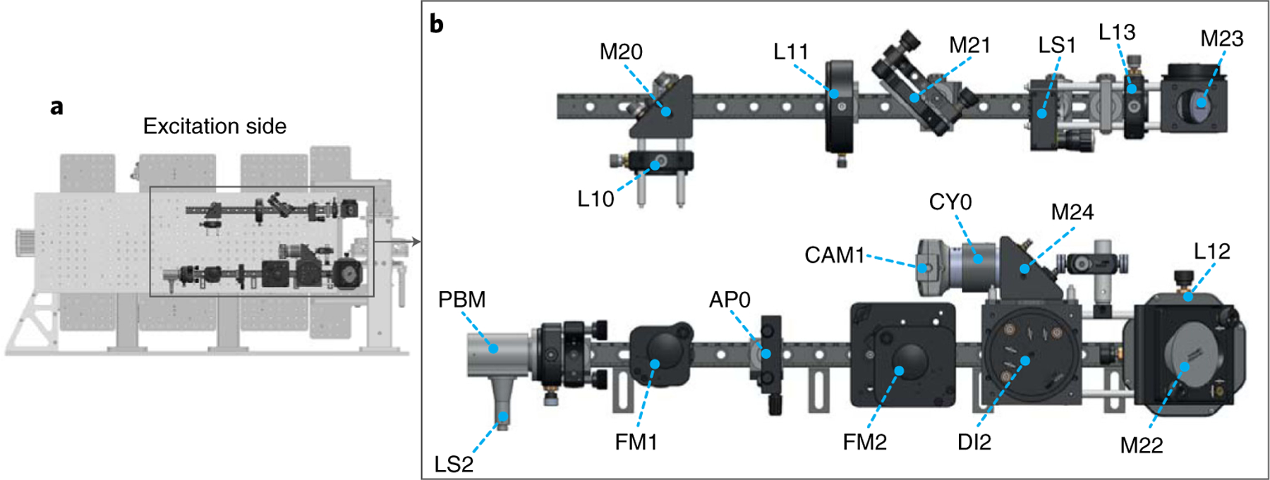
The development of single-molecule switching (SMS) fluorescence microscopy (also called single-molecule localization microscopy) over the last decade has enabled researchers to image cell biological structures at unprecedented resolution. Using two opposing objectives in a so-called 4Pi geometry doubles the available numerical aperture, and coupling this with interferometric detection has demonstrated 3D resolution down to 10 nm over entire cellular volumes. The aim of this protocol is to enable interested researchers to establish 4Pi-SMS super-resolution microscopy in their laboratories. We describe in detail how to assemble the optomechanical components of a 4Pi-SMS instrument, align its optical beampath and test its performance. The protocol further provides instructions on how to prepare test samples of fluorescent beads, operate this instrument to acquire images of whole cells and analyze the raw image data to reconstruct super-resolution 3D data sets. Furthermore, we provide a troubleshooting guide and present examples of anticipated results. An experienced optical instrument builder will require ~12 months from the start of ordering hardware components to acquiring high-quality biological images.

摘要

过去十年中,单分子荧光显微镜(也称为单分子定位显微镜)的发展使研究人员能够以前所未有的分辨率对细胞生物结构进行成像。在所谓的 4Pi 几何结构中使用两个相对的物镜可以将可用的数值孔径增加一倍,并且将其与干涉检测相结合,已经证明在整个细胞体积中可以达到 3D 分辨率低至 10nm。本协议的目的是使有兴趣的研究人员能够在他们的实验室中建立 4Pi-SMS 超分辨率显微镜。我们详细描述了如何组装 4Pi-SMS 仪器的光机组件,对准其光学光路并测试其性能。该协议还提供了有关如何制备荧光珠测试样品、操作该仪器以获取整个细胞图像以及分析原始图像数据以重建超分辨率 3D 数据集的说明。此外,我们提供了故障排除指南,并展示了预期结果的示例。有经验的光学仪器制造商从开始订购硬件组件到获得高质量的生物图像,大约需要 12 个月的时间。

https://cdn.ncbi.nlm.nih.gov/pmc/blobs/3a1f/9118368/3d0adde545cb/nihms-1803679-f0026.jpg
https://cdn.ncbi.nlm.nih.gov/pmc/blobs/3a1f/9118368/236e3ad4c9cc/nihms-1803679-f0002.jpg
https://cdn.ncbi.nlm.nih.gov/pmc/blobs/3a1f/9118368/b5eb2ca7d9c4/nihms-1803679-f0003.jpg
https://cdn.ncbi.nlm.nih.gov/pmc/blobs/3a1f/9118368/14fef094dd68/nihms-1803679-f0004.jpg
https://cdn.ncbi.nlm.nih.gov/pmc/blobs/3a1f/9118368/046af6b4b6a9/nihms-1803679-f0005.jpg
https://cdn.ncbi.nlm.nih.gov/pmc/blobs/3a1f/9118368/887bc9f58cba/nihms-1803679-f0006.jpg
https://cdn.ncbi.nlm.nih.gov/pmc/blobs/3a1f/9118368/c05154bf6805/nihms-1803679-f0007.jpg
https://cdn.ncbi.nlm.nih.gov/pmc/blobs/3a1f/9118368/0d229d1bcd63/nihms-1803679-f0008.jpg
https://cdn.ncbi.nlm.nih.gov/pmc/blobs/3a1f/9118368/0ba56cd313dc/nihms-1803679-f0009.jpg
https://cdn.ncbi.nlm.nih.gov/pmc/blobs/3a1f/9118368/89ae966da804/nihms-1803679-f0010.jpg
https://cdn.ncbi.nlm.nih.gov/pmc/blobs/3a1f/9118368/cf19d1cea683/nihms-1803679-f0011.jpg
https://cdn.ncbi.nlm.nih.gov/pmc/blobs/3a1f/9118368/434f0b4db051/nihms-1803679-f0012.jpg
https://cdn.ncbi.nlm.nih.gov/pmc/blobs/3a1f/9118368/9c5e6d6b856e/nihms-1803679-f0013.jpg
https://cdn.ncbi.nlm.nih.gov/pmc/blobs/3a1f/9118368/3644f4d0603a/nihms-1803679-f0014.jpg
https://cdn.ncbi.nlm.nih.gov/pmc/blobs/3a1f/9118368/58d6fb0214b1/nihms-1803679-f0015.jpg
https://cdn.ncbi.nlm.nih.gov/pmc/blobs/3a1f/9118368/d66a3402358c/nihms-1803679-f0016.jpg
https://cdn.ncbi.nlm.nih.gov/pmc/blobs/3a1f/9118368/62ae9fc18e68/nihms-1803679-f0017.jpg
https://cdn.ncbi.nlm.nih.gov/pmc/blobs/3a1f/9118368/a01b70be8a31/nihms-1803679-f0018.jpg
https://cdn.ncbi.nlm.nih.gov/pmc/blobs/3a1f/9118368/2c7fb76c1988/nihms-1803679-f0019.jpg
https://cdn.ncbi.nlm.nih.gov/pmc/blobs/3a1f/9118368/7a9907150309/nihms-1803679-f0020.jpg
https://cdn.ncbi.nlm.nih.gov/pmc/blobs/3a1f/9118368/dd1f182b84b2/nihms-1803679-f0021.jpg
https://cdn.ncbi.nlm.nih.gov/pmc/blobs/3a1f/9118368/719c066a59b3/nihms-1803679-f0022.jpg
https://cdn.ncbi.nlm.nih.gov/pmc/blobs/3a1f/9118368/431d2bfa2e9c/nihms-1803679-f0023.jpg
https://cdn.ncbi.nlm.nih.gov/pmc/blobs/3a1f/9118368/a049443a966a/nihms-1803679-f0024.jpg
https://cdn.ncbi.nlm.nih.gov/pmc/blobs/3a1f/9118368/446dfd9487d7/nihms-1803679-f0025.jpg
https://cdn.ncbi.nlm.nih.gov/pmc/blobs/3a1f/9118368/3d0adde545cb/nihms-1803679-f0026.jpg

相似文献

1
Implementation of a 4Pi-SMS super-resolution microscope.4Pi-SMS 超分辨率显微镜的实现。
Nat Protoc. 2021 Feb;16(2):677-727. doi: 10.1038/s41596-020-00428-7. Epub 2020 Dec 16.
2
Build and operation of a custom 3D, multicolor, single-molecule localization microscope.定制的三维、多色、单分子定位显微镜的构建与操作。
Nat Protoc. 2024 Aug;19(8):2467-2525. doi: 10.1038/s41596-024-00989-x. Epub 2024 May 3.
3
Enhanced 4Pi single-molecule localization microscopy with coherent pupil based localization.基于相干光瞳定位的增强型 4Pi 单分子定位显微镜
Commun Biol. 2020 May 8;3(1):220. doi: 10.1038/s42003-020-0908-2.
4
Ultra-High Resolution 3D Imaging of Whole Cells.全细胞的超高分辨率3D成像。
Cell. 2016 Aug 11;166(4):1028-1040. doi: 10.1016/j.cell.2016.06.016. Epub 2016 Jul 7.
5
Chemistry of Photosensitive Fluorophores for Single-Molecule Localization Microscopy.用于单分子定位显微镜的光敏荧光团化学
ACS Chem Biol. 2019 Jun 21;14(6):1077-1090. doi: 10.1021/acschembio.9b00197. Epub 2019 May 13.
6
Building a super-resolution fluorescence cryomicroscope.构建超分辨率荧光冷冻显微镜。
Methods Cell Biol. 2024;187:205-222. doi: 10.1016/bs.mcb.2024.02.026. Epub 2024 Mar 13.
7
3D reconstruction of high-resolution STED microscope images.高分辨率受激发射损耗(STED)显微镜图像的三维重建
Microsc Res Tech. 2008 Sep;71(9):644-50. doi: 10.1002/jemt.20602.
8
Correlative super-resolution fluorescence and electron microscopy using conventional fluorescent proteins in vacuo.在真空中使用传统荧光蛋白的相关超分辨率荧光和电子显微镜技术。
J Struct Biol. 2017 Aug;199(2):120-131. doi: 10.1016/j.jsb.2017.05.013. Epub 2017 May 30.
9
Super-Resolution Microscopy Techniques and Their Potential for Applications in Radiation Biophysics.超分辨率显微镜技术及其在辐射生物物理学中的应用潜力
Methods Mol Biol. 2017;1663:1-13. doi: 10.1007/978-1-4939-7265-4_1.
10
Multiplexed 3D super-resolution imaging of whole cells using spinning disk confocal microscopy and DNA-PAINT.基于旋转盘共聚焦显微镜和 DNA-PAINT 的全细胞多重 3D 超分辨率成像
Nat Commun. 2017 Dec 12;8(1):2090. doi: 10.1038/s41467-017-02028-8.

引用本文的文献

1
Cellular optical imaging techniques: a dynamic advancing frontier.细胞光学成像技术:一个动态发展的前沿领域。
Sci China Life Sci. 2025 Jul 16. doi: 10.1007/s11427-024-2916-5.
2
Immobile lipopolysaccharides and outer membrane proteins differentially segregate in growing .固定不动的脂多糖和外膜蛋白在生长过程中差异分离。
Proc Natl Acad Sci U S A. 2025 Mar 11;122(10):e2414725122. doi: 10.1073/pnas.2414725122. Epub 2025 Mar 3.
3
MINFLUX Nanoscopy: A "Brilliant" Technique Promising Major Breakthrough.MINFLUX纳米显微镜:一项有望取得重大突破的“卓越”技术。
Microsc Res Tech. 2025 May;88(5):1264-1272. doi: 10.1002/jemt.24765. Epub 2025 Jan 21.
4
A tactile perception method with flexible grating structural color.一种具有柔性光栅结构色的触觉感知方法。
Natl Sci Rev. 2024 Nov 15;12(1):nwae413. doi: 10.1093/nsr/nwae413. eCollection 2025 Jan.
5
Elucidating subcellular architecture and dynamics at isotropic 100-nm resolution with 4Pi-SIM.利用4Pi-SIM以各向同性100纳米分辨率阐明亚细胞结构和动力学。
Nat Methods. 2025 Feb;22(2):335-347. doi: 10.1038/s41592-024-02515-z. Epub 2024 Dec 23.
6
VPS13B is localized at the interface between Golgi cisternae and is a functional partner of FAM177A1.VPS13B 位于高尔基体潴腔的界面处,是 FAM177A1 的功能伙伴。
J Cell Biol. 2024 Dec 2;223(12). doi: 10.1083/jcb.202311189. Epub 2024 Sep 27.
7
Universal inverse modeling of point spread functions for SMLM localization and microscope characterization.用于 SMLM 定位和显微镜特性分析的点扩散函数的通用逆建模。
Nat Methods. 2024 Jun;21(6):1082-1093. doi: 10.1038/s41592-024-02282-x. Epub 2024 Jun 3.
8
Advancements and Practical Considerations for Biophysical Research: Navigating the Challenges and Future of Super-resolution Microscopy.生物物理研究的进展与实际考量:应对超分辨率显微镜的挑战与未来
Chem Biomed Imaging. 2024 Apr 19;2(5):331-344. doi: 10.1021/cbmi.4c00019. eCollection 2024 May 27.
9
Dynamics and functions of E-cadherin complexes in epithelial cell and tissue morphogenesis.上皮细胞和组织形态发生过程中E-钙黏蛋白复合体的动力学与功能
Mar Life Sci Technol. 2023 Nov 24;5(4):585-601. doi: 10.1007/s42995-023-00206-w. eCollection 2023 Nov.
10
Universal inverse modelling of point spread functions for SMLM localization and microscope characterization.用于超分辨显微镜定位和显微镜表征的点扩散函数通用逆建模
bioRxiv. 2023 Oct 26:2023.10.26.564064. doi: 10.1101/2023.10.26.564064.