Translational Neuromodeling Unit, University of Zurich and ETH Zurich, Zurich, Switzerland.
Hospital of Psychiatry, University of Zurich, Zurich, Switzerland.
Sci Rep. 2020 Dec 18;10(1):22346. doi: 10.1038/s41598-020-79170-9.
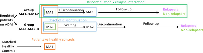
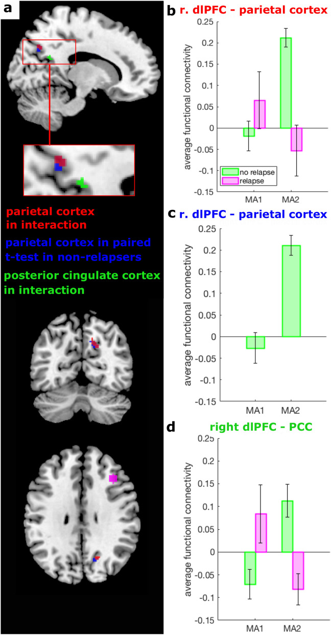
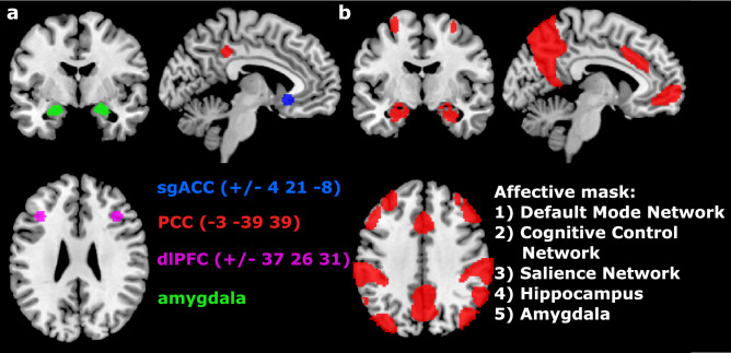
The risk of relapsing into depression after stopping antidepressants is high, but no established predictors exist. Resting-state functional magnetic resonance imaging (rsfMRI) measures may help predict relapse and identify the mechanisms by which relapses occur. rsfMRI data were acquired from healthy controls and from patients with remitted major depressive disorder on antidepressants. Patients were assessed a second time either before or after discontinuation of the antidepressant, and followed up for six months to assess relapse. A seed-based functional connectivity analysis was conducted focusing on the left subgenual anterior cingulate cortex and left posterior cingulate cortex. Seeds in the amygdala and dorsolateral prefrontal cortex were explored. 44 healthy controls (age: 33.8 (10.5), 73% female) and 84 patients (age: 34.23 (10.8), 80% female) were included in the analysis. 29 patients went on to relapse and 38 remained well. The seed-based analysis showed that discontinuation resulted in an increased functional connectivity between the right dorsolateral prefrontal cortex and the parietal cortex in non-relapsers. In an exploratory analysis, this functional connectivity predicted relapse risk with a balanced accuracy of 0.86. Further seed-based analyses, however, failed to reveal differences in functional connectivity between patients and controls, between relapsers and non-relapsers before discontinuation and changes due to discontinuation independent of relapse. In conclusion, changes in the connectivity between the dorsolateral prefrontal cortex and the posterior default mode network were associated with and predictive of relapse after open-label antidepressant discontinuation. This finding requires replication in a larger dataset.
抗抑郁药停药后复发抑郁的风险很高,但目前尚无确定的预测指标。静息态功能磁共振成像(rsfMRI)测量可能有助于预测复发,并确定复发发生的机制。rsfMRI 数据来自服用抗抑郁药的缓解期重度抑郁症患者和健康对照者。患者在停药前或停药后进行第二次评估,并随访 6 个月以评估复发情况。进行了基于种子的功能连接分析,重点关注左侧前扣带回皮质亚区和左侧后扣带回皮质。还探索了杏仁核和背外侧前额叶皮层的种子。纳入了 44 名健康对照者(年龄:33.8(10.5),73%为女性)和 84 名患者(年龄:34.23(10.8),80%为女性)进行分析。29 名患者复发,38 名患者病情稳定。基于种子的分析显示,在未复发者中,停药后右侧背外侧前额叶皮层与顶叶皮层之间的功能连接增加。在一项探索性分析中,这种功能连接预测复发风险的准确率为 0.86。然而,进一步的基于种子的分析未能显示出患者与对照组、停药前复发者与非复发者之间的功能连接差异,以及与复发无关的停药后变化。总之,背外侧前额叶皮层与后默认模式网络之间连接的变化与停药后开放性抗抑郁药停药后的复发相关,并具有预测作用。这一发现需要在更大的数据集上进行复制。