• 文献检索
  • 文档翻译
  • 深度研究
  • 学术资讯
  • Suppr Zotero 插件Zotero 插件
  • 邀请有礼
  • 套餐&价格
  • 历史记录
应用&插件
Suppr Zotero 插件Zotero 插件浏览器插件Mac 客户端Windows 客户端微信小程序
定价
高级版会员购买积分包购买API积分包
服务
文献检索文档翻译深度研究API 文档MCP 服务
关于我们
关于 Suppr公司介绍联系我们用户协议隐私条款
关注我们

Suppr 超能文献

核心技术专利:CN118964589B侵权必究
粤ICP备2023148730 号-1Suppr @ 2026

文献检索

告别复杂PubMed语法,用中文像聊天一样搜索,搜遍4000万医学文献。AI智能推荐,让科研检索更轻松。

立即免费搜索

文件翻译

保留排版,准确专业,支持PDF/Word/PPT等文件格式,支持 12+语言互译。

免费翻译文档

深度研究

AI帮你快速写综述,25分钟生成高质量综述,智能提取关键信息,辅助科研写作。

立即免费体验

基于皮质-皮质诱发电位的癫痫发作起始区定位的传递函数模型

Transfer Function Models for the Localization of Seizure Onset Zone From Cortico-Cortical Evoked Potentials.

作者信息

Kamali Golnoosh, Smith Rachel June, Hays Mark, Coogan Christopher, Crone Nathan E, Kang Joon Y, Sarma Sridevi V

机构信息

Neuromedical Control Systems Laboratory, Department of Electrical and Computer Engineering, Institute of Computational Medicine, Johns Hopkins University, Baltimore, MD, United States.

Neuromedical Control Systems Laboratory, Department of Biomedical Engineering, Institute of Computational Medicine, Johns Hopkins University, Baltimore, MD, United States.

出版信息

Front Neurol. 2020 Dec 10;11:579961. doi: 10.3389/fneur.2020.579961. eCollection 2020.

DOI:10.3389/fneur.2020.579961
PMID:33362689
原文链接:https://pmc.ncbi.nlm.nih.gov/articles/PMC7758451/
Abstract

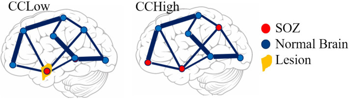
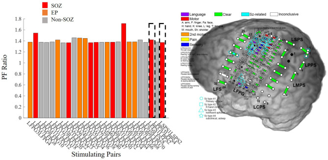
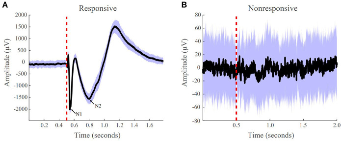
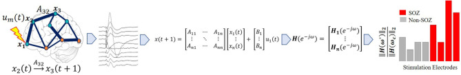
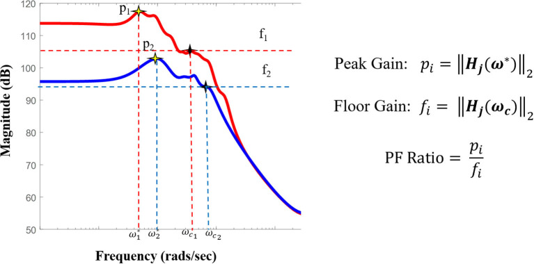
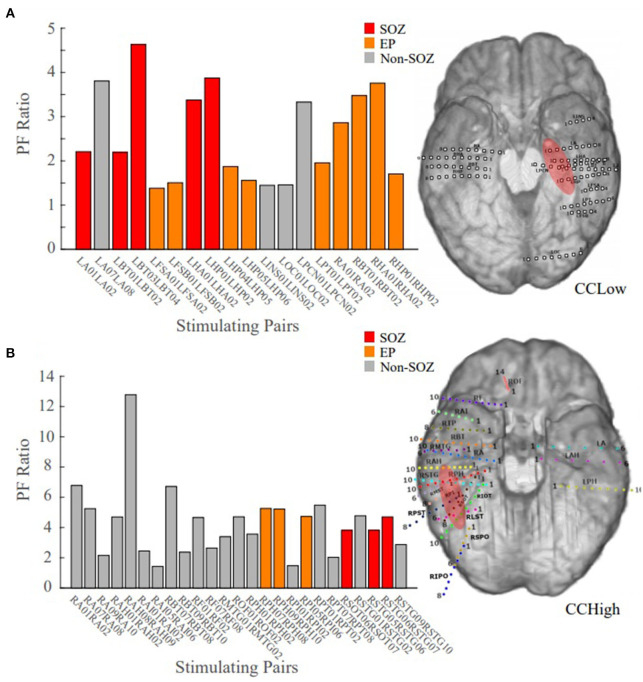
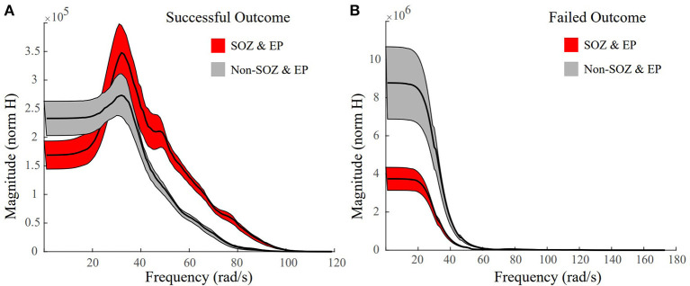
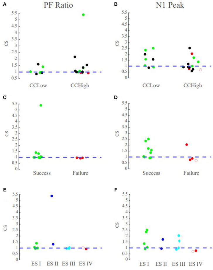
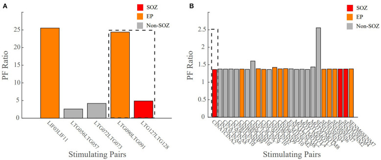
Surgical resection of the seizure onset zone (SOZ) could potentially lead to seizure-freedom in medically refractory epilepsy patients. However, localizing the SOZ can be a time consuming and tedious process involving visual inspection of intracranial electroencephalographic (iEEG) recordings captured during passive patient monitoring. Cortical stimulation is currently performed on patients undergoing invasive EEG monitoring for the main purpose of mapping functional brain networks such as language and motor networks. We hypothesized that evoked responses from single pulse electrical stimulation (SPES) can also be used to localize the SOZ as they may express the natural frequencies and connectivity of the iEEG network. To test our hypothesis, we constructed patient specific transfer function models from the evoked responses recorded from 22 epilepsy patients that underwent SPES evaluation and iEEG monitoring. We then computed the frequency and connectivity dependent "peak gain" of the system as measured by the norm from systems theory. We found that in cases for which clinicians had high confidence in localizing the SOZ, the highest peak gain transfer functions with the smallest "floor gain" (gain at which the dipped 3dB below DC gain) corresponded to when the clinically annotated SOZ and early spread regions were stimulated. In more complex cases, there was a large spread of the peak-to-floor (PF) ratios when the clinically annotated SOZ was stimulated. Interestingly for patients who had successful surgeries, our ratio of gains, agreed with clinical localization, no matter the complexity of the case. For patients with failed surgeries, the PF ratio did not match clinical annotations. Our findings suggest that transfer function gains and their corresponding frequency responses computed from SPES evoked responses may improve SOZ localization and thus surgical outcomes.

摘要

手术切除癫痫发作起始区(SOZ)可能会使药物难治性癫痫患者实现无癫痫发作。然而,定位SOZ可能是一个耗时且繁琐的过程,需要对被动患者监测期间捕获的颅内脑电图(iEEG)记录进行目视检查。目前,皮质刺激是在接受侵入性脑电图监测的患者身上进行的,主要目的是绘制功能性脑网络,如语言和运动网络。我们假设单脉冲电刺激(SPES)诱发的反应也可用于定位SOZ,因为它们可能表达了iEEG网络的固有频率和连通性。为了验证我们的假设,我们根据22例接受SPES评估和iEEG监测的癫痫患者记录的诱发反应构建了患者特异性传递函数模型。然后,我们根据系统理论中的范数计算了系统的频率和连通性相关的“峰值增益”。我们发现,在临床医生对SOZ定位有高度信心的病例中,具有最小“底增益”(比直流增益低3dB时的增益)的最高峰值增益传递函数对应于刺激临床标注的SOZ和早期扩散区域时。在更复杂的病例中,刺激临床标注的SOZ时,峰底(PF)比的分布范围很大。有趣的是,对于手术成功的患者,无论病例的复杂性如何,我们的增益比都与临床定位一致。对于手术失败的患者,PF比与临床标注不匹配。我们的研究结果表明,从SPES诱发反应计算得到的传递函数增益及其相应的频率响应可能会改善SOZ定位,从而改善手术结果。

https://cdn.ncbi.nlm.nih.gov/pmc/blobs/c306/7758451/f9a001c99d20/fneur-11-579961-g0010.jpg
https://cdn.ncbi.nlm.nih.gov/pmc/blobs/c306/7758451/e7f58d4370c2/fneur-11-579961-g0001.jpg
https://cdn.ncbi.nlm.nih.gov/pmc/blobs/c306/7758451/f46a24e1f3ad/fneur-11-579961-g0002.jpg
https://cdn.ncbi.nlm.nih.gov/pmc/blobs/c306/7758451/311483c11079/fneur-11-579961-g0003.jpg
https://cdn.ncbi.nlm.nih.gov/pmc/blobs/c306/7758451/1d5144b39214/fneur-11-579961-g0004.jpg
https://cdn.ncbi.nlm.nih.gov/pmc/blobs/c306/7758451/4f823db2d9b9/fneur-11-579961-g0005.jpg
https://cdn.ncbi.nlm.nih.gov/pmc/blobs/c306/7758451/0df52f035754/fneur-11-579961-g0006.jpg
https://cdn.ncbi.nlm.nih.gov/pmc/blobs/c306/7758451/72a242df6050/fneur-11-579961-g0007.jpg
https://cdn.ncbi.nlm.nih.gov/pmc/blobs/c306/7758451/934d11bbbae1/fneur-11-579961-g0008.jpg
https://cdn.ncbi.nlm.nih.gov/pmc/blobs/c306/7758451/4d40c05bfb8a/fneur-11-579961-g0009.jpg
https://cdn.ncbi.nlm.nih.gov/pmc/blobs/c306/7758451/f9a001c99d20/fneur-11-579961-g0010.jpg

相似文献

1
Transfer Function Models for the Localization of Seizure Onset Zone From Cortico-Cortical Evoked Potentials.基于皮质-皮质诱发电位的癫痫发作起始区定位的传递函数模型
Front Neurol. 2020 Dec 10;11:579961. doi: 10.3389/fneur.2020.579961. eCollection 2020.
2
Localizing the seizure onset zone from single pulse electrical stimulation responses using transfer function models.使用传递函数模型从单脉冲电刺激反应中定位癫痫发作起始区。
Annu Int Conf IEEE Eng Med Biol Soc. 2020 Jul;2020:2524-2527. doi: 10.1109/EMBC44109.2020.9175954.
3
State-space models of evoked potentials to localize the seizure onset zone.用于定位癫痫发作起始区的诱发电位状态空间模型。
Annu Int Conf IEEE Eng Med Biol Soc. 2020 Jul;2020:2528-2531. doi: 10.1109/EMBC44109.2020.9176697.
4
Network excitability of stimulation-induced spectral responses helps localize the seizure onset zone.刺激诱导的光谱反应的网络兴奋性有助于定位癫痫发作起始区。
Clin Neurophysiol. 2024 Oct;166:43-55. doi: 10.1016/j.clinph.2024.07.010. Epub 2024 Jul 24.
5
Cortico-cortical evoked potentials in response to varying stimulation intensity improves seizure localization.皮层-皮层诱发电位对不同刺激强度的反应可改善癫痫定位。
Clin Neurophysiol. 2023 Jan;145:119-128. doi: 10.1016/j.clinph.2022.08.024. Epub 2022 Sep 11.
6
Stimulating native seizures with neural resonance: a new approach to localize the seizure onset zone.用神经共振刺激原生发作:一种定位发作起始区的新方法。
Brain. 2022 Nov 21;145(11):3886-3900. doi: 10.1093/brain/awac214.
7
The effectiveness of cortico-cortical evoked potential in detecting seizure onset zones.皮质-皮质诱发电位在检测癫痫发作起始区中的有效性。
Neurol Res. 2018 Jun;40(6):480-490. doi: 10.1080/01616412.2018.1454092. Epub 2018 Mar 24.
8
Transient seizure onset network for localization of epileptogenic zone: effective connectivity and graph theory-based analyses of ECoG data in temporal lobe epilepsy.短暂性发作起始网络用于致痫灶定位:基于 ECoG 数据的有效连通性和图论分析在颞叶癫痫中的应用。
J Neurol. 2019 Apr;266(4):844-859. doi: 10.1007/s00415-019-09204-4. Epub 2019 Jan 25.
9
High frequency activity overriding cortico-cortical evoked potentials reflects altered excitability in the human epileptic focus.高频活动凌驾于皮质-皮质诱发电位之上反映了人类癫痫病灶兴奋性的改变。
Clin Neurophysiol. 2017 Sep;128(9):1673-1681. doi: 10.1016/j.clinph.2017.06.249. Epub 2017 Jul 8.
10
Ictal and interictal source imaging on intracranial EEG predicts epilepsy surgery outcome in children with focal cortical dysplasia.颅内 EEG 的发作期和发作间期源成像预测局灶性皮质发育不良儿童的癫痫手术结果。
Clin Neurophysiol. 2020 Mar;131(3):734-743. doi: 10.1016/j.clinph.2019.12.408. Epub 2020 Jan 20.

引用本文的文献

1
Effective Connectivity Predicts Surgical Outcomes in Temporal Lobe Epilepsy: A SEEG Study.有效连接性可预测颞叶癫痫的手术结果:一项立体定向脑电图研究
CNS Neurosci Ther. 2025 Aug;31(8):e70563. doi: 10.1111/cns.70563.
2
Exploring synergies: Advancing neuroscience with machine learning.探索协同效应:借助机器学习推动神经科学发展。
Signal Processing. 2026 Jan;238. doi: 10.1016/j.sigpro.2025.110116. Epub 2025 Jun 2.
3
The sixth sense: how much does interictal intracranial EEG add to determining the focality of epileptic networks?第六感:发作间期颅内脑电图在确定癫痫网络的局灶性方面能增加多少价值?

本文引用的文献

1
(Re)Defining success in epilepsy surgery: The importance of relative seizure reduction in patient-reported quality of life.(重新)定义癫痫手术的成功:患者报告的生活质量中相对发作减少的重要性。
Epilepsia. 2019 Oct;60(10):2078-2085. doi: 10.1111/epi.16327. Epub 2019 Aug 28.
2
Connectivity of the human insula: A cortico-cortical evoked potential (CCEP) study.人脑岛的连接:皮质-皮质诱发电位(CCEP)研究。
Cortex. 2019 Nov;120:419-442. doi: 10.1016/j.cortex.2019.05.019. Epub 2019 Jun 18.
3
Application of Graph Theory for Identifying Connectivity Patterns in Human Brain Networks: A Systematic Review.
Brain Commun. 2024 Sep 27;6(5):fcae320. doi: 10.1093/braincomms/fcae320. eCollection 2024.
4
Virtual stimulation of the interictal EEG network localizes the EZ as a measure of cortical excitability.发作间期脑电图网络的虚拟刺激将致痫区定位为皮质兴奋性的一种测量指标。
Front Netw Physiol. 2024 Aug 20;4:1425625. doi: 10.3389/fnetp.2024.1425625. eCollection 2024.
5
Network excitability of stimulation-induced spectral responses helps localize the seizure onset zone.刺激诱导的光谱反应的网络兴奋性有助于定位癫痫发作起始区。
Clin Neurophysiol. 2024 Oct;166:43-55. doi: 10.1016/j.clinph.2024.07.010. Epub 2024 Jul 24.
6
Quantifying interictal intracranial EEG to predict focal epilepsy.量化发作间期颅内脑电图以预测局灶性癫痫。
ArXiv. 2023 Jul 27:arXiv:2307.15170v1.
7
Stimulation to probe, excite, and inhibit the epileptic brain.刺激探针,激发和抑制癫痫大脑。
Epilepsia. 2023 Dec;64 Suppl 3(Suppl 3):S49-S61. doi: 10.1111/epi.17640. Epub 2023 May 18.
8
Passive and active markers of cortical excitability in epilepsy.癫痫的皮质兴奋性的被动和主动标志物。
Epilepsia. 2023 Dec;64 Suppl 3(Suppl 3):S25-S36. doi: 10.1111/epi.17578. Epub 2023 Mar 22.
9
Quantifying trial-by-trial variability during cortico-cortical evoked potential mapping of epileptogenic tissue.量化癫痫组织皮质皮质诱发电位图的试验间变异性。
Epilepsia. 2023 Apr;64(4):1021-1034. doi: 10.1111/epi.17528. Epub 2023 Feb 22.
10
Quantitative approaches to guide epilepsy surgery from intracranial EEG.从颅内脑电图引导癫痫手术的定量方法。
Brain. 2023 Jun 1;146(6):2248-2258. doi: 10.1093/brain/awad007.
图论在识别人类脑网络连接模式中的应用:一项系统综述。
Front Neurosci. 2019 Jun 6;13:585. doi: 10.3389/fnins.2019.00585. eCollection 2019.
4
Localization of Epileptogenic Zone Based on Cortico-Cortical Evoked Potential (CCEP): A Feature Extraction and Graph Theory Approach.基于皮质-皮质诱发电位(CCEP)的癫痫发作起始区定位:一种特征提取与图论方法
Front Neuroinform. 2019 Apr 24;13:31. doi: 10.3389/fninf.2019.00031. eCollection 2019.
5
Delayed high-frequency suppression after automated single-pulse electrical stimulation identifies the seizure onset zone in patients with refractory epilepsy.自动单脉冲电刺激后的迟发性高频抑制可确定耐药性癫痫患者的癫痫发作起始区。
Clin Neurophysiol. 2018 Nov;129(11):2466-2474. doi: 10.1016/j.clinph.2018.06.021. Epub 2018 Jul 4.
6
Evoked directional network characteristics of epileptogenic tissue derived from single pulse electrical stimulation.单脉冲电刺激致痫性组织的诱导向量网络特征。
Hum Brain Mapp. 2018 Nov;39(11):4611-4622. doi: 10.1002/hbm.24309. Epub 2018 Jul 21.
7
The effectiveness of cortico-cortical evoked potential in detecting seizure onset zones.皮质-皮质诱发电位在检测癫痫发作起始区中的有效性。
Neurol Res. 2018 Jun;40(6):480-490. doi: 10.1080/01616412.2018.1454092. Epub 2018 Mar 24.
8
Getting the best outcomes from epilepsy surgery.从癫痫手术中获得最佳结果。
Ann Neurol. 2018 Apr;83(4):676-690. doi: 10.1002/ana.25205. Epub 2018 Apr 10.
9
The Hippocampus and Amygdala Are Integrators of Neocortical Influence: A CorticoCortical Evoked Potential Study.海马体和杏仁核是新皮层影响的整合者:皮质皮质诱发电位研究。
Brain Connect. 2017 Dec;7(10):648-660. doi: 10.1089/brain.2017.0527.
10
High frequency activity overriding cortico-cortical evoked potentials reflects altered excitability in the human epileptic focus.高频活动凌驾于皮质-皮质诱发电位之上反映了人类癫痫病灶兴奋性的改变。
Clin Neurophysiol. 2017 Sep;128(9):1673-1681. doi: 10.1016/j.clinph.2017.06.249. Epub 2017 Jul 8.