Kim Yu Rim, Kim Young Min, Lee Jaeho, Park Joohyun, Lee Jong Eun, Hyun Young-Min
Department of Anatomy, Yonsei University College of Medicine, Seoul, South Korea.
BK21 PLUS Project for Medical Science, Yonsei University College of Medicine, Seoul, South Korea.
Front Cell Dev Biol. 2020 Dec 8;8:613733. doi: 10.3389/fcell.2020.613733. eCollection 2020.
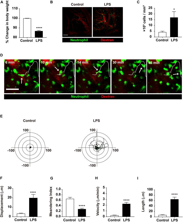
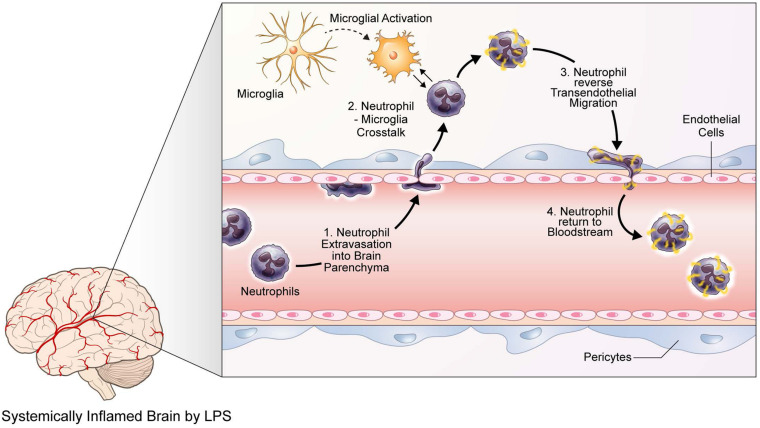
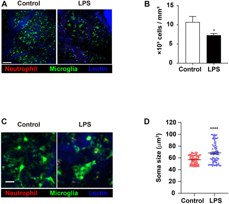
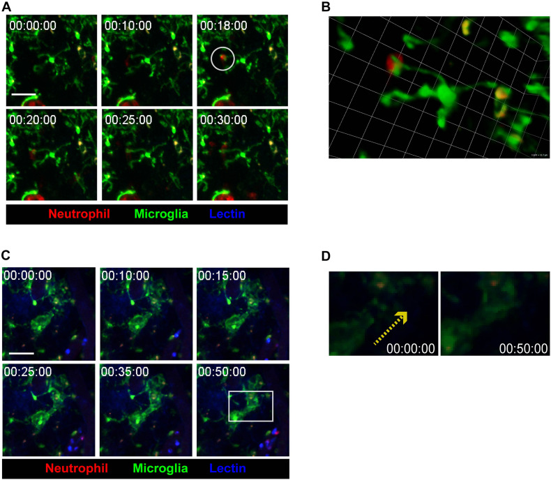
The circulatory neutrophil and brain tissue-resident microglia are two important immune cells involved in neuroinflammation. Since neutrophils that infiltrate through the brain vascular vessel may affect the immune function of microglia in the brain, close investigation of the interaction between these cells is important in understanding neuroinflammatory phenomena and immunological aftermaths that follow. This study aimed to observe how morphology and function of both neutrophils and microglia are converted in the inflamed brain. To directly investigate cellular responses of neutrophils and microglia, LysM and CXCR1 mice were used for the observation of neutrophils and microglia, respectively. In addition, low-dose lipopolysaccharide (LPS) was utilized to induce acute inflammation in the central nervous system (CNS) of mice. Real-time observation on mice brain undergoing neuroinflammation via two-photon intravital microscopy revealed various changes in neutrophils and microglia; namely, neutrophil infiltration and movement within the brain tissue increased, while microglia displayed morphological changes suggesting an activated state. Furthermore, neutrophils seemed to not only actively interact with microglial processes but also exhibit reverse transendothelial migration (rTEM) back to the bloodstream. Thus, it may be postulated that, through crosstalk with neutrophils, macrophages are primed to initiate a neuroinflammatory immune response; also, during pathogenic events in the brain, neutrophils that engage in rTEM may deliver proinflammatory signals to peripheral organs outside the brain. Taken together, these results both show that neuroinflammation results in significant alterations in neutrophils and microglia and lay the pavement for further studies on the molecular mechanisms behind such changes.
循环中的中性粒细胞和脑组织驻留的小胶质细胞是参与神经炎症的两种重要免疫细胞。由于通过脑血管浸润的中性粒细胞可能会影响脑中的小胶质细胞免疫功能,因此密切研究这些细胞之间的相互作用对于理解随后发生的神经炎症现象和免疫后果至关重要。本研究旨在观察在炎症状态下脑中中性粒细胞和小胶质细胞的形态及功能是如何转变的。为了直接研究中性粒细胞和小胶质细胞的细胞反应,分别使用LysM和CXCR1小鼠来观察中性粒细胞和小胶质细胞。此外,利用低剂量脂多糖(LPS)诱导小鼠中枢神经系统(CNS)发生急性炎症。通过双光子活体显微镜对经历神经炎症的小鼠大脑进行实时观察,发现中性粒细胞和小胶质细胞发生了各种变化;具体而言,脑组织内中性粒细胞的浸润和移动增加,而小胶质细胞则呈现出提示激活状态的形态变化。此外,中性粒细胞似乎不仅与小胶质细胞的突起积极相互作用,还表现出反向跨内皮迁移(rTEM)回到血流中。因此,可以推测,通过与中性粒细胞的相互作用,巨噬细胞被激活以启动神经炎症免疫反应;而且,在脑部发生致病事件期间,进行rTEM的中性粒细胞可能会将促炎信号传递到脑外的外周器官。综上所述,这些结果表明神经炎症会导致中性粒细胞和小胶质细胞发生显著改变,并为进一步研究这些变化背后的分子机制奠定了基础。