• 文献检索
  • 文档翻译
  • 深度研究
  • 学术资讯
  • Suppr Zotero 插件Zotero 插件
  • 邀请有礼
  • 套餐&价格
  • 历史记录
应用&插件
Suppr Zotero 插件Zotero 插件浏览器插件Mac 客户端Windows 客户端微信小程序
定价
高级版会员购买积分包购买API积分包
服务
文献检索文档翻译深度研究API 文档MCP 服务
关于我们
关于 Suppr公司介绍联系我们用户协议隐私条款
关注我们

Suppr 超能文献

核心技术专利:CN118964589B侵权必究
粤ICP备2023148730 号-1Suppr @ 2026

文献检索

告别复杂PubMed语法,用中文像聊天一样搜索,搜遍4000万医学文献。AI智能推荐,让科研检索更轻松。

立即免费搜索

文件翻译

保留排版,准确专业,支持PDF/Word/PPT等文件格式,支持 12+语言互译。

免费翻译文档

深度研究

AI帮你快速写综述,25分钟生成高质量综述,智能提取关键信息,辅助科研写作。

立即免费体验

用于骨软骨修复和关节表面置换的一体化双相解剖间充质基质细胞负载复合支架的制造和成熟。

Fabrication and maturation of integrated biphasic anatomic mesenchymal stromal cell-laden composite scaffolds for osteochondral repair and joint resurfacing.

机构信息

McKay Orthopaedic Research Laboratory, University of Pennsylvania, Philadelphia, Pennsylvania, USA.

Translational Musculoskeletal Research Center, Philadelphia VA Medical Center, Philadelphia, Pennsylvania, USA.

出版信息

J Orthop Res. 2021 Nov;39(11):2323-2332. doi: 10.1002/jor.24969. Epub 2021 Jan 14.

DOI:10.1002/jor.24969
PMID:33368606
原文链接:https://pmc.ncbi.nlm.nih.gov/articles/PMC8222412/
Abstract

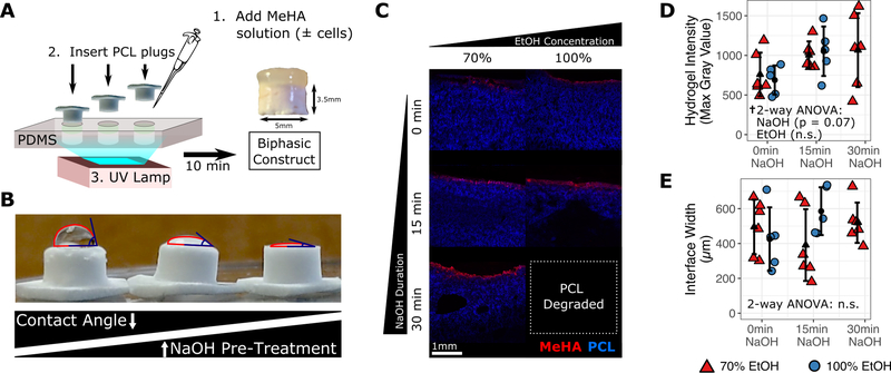
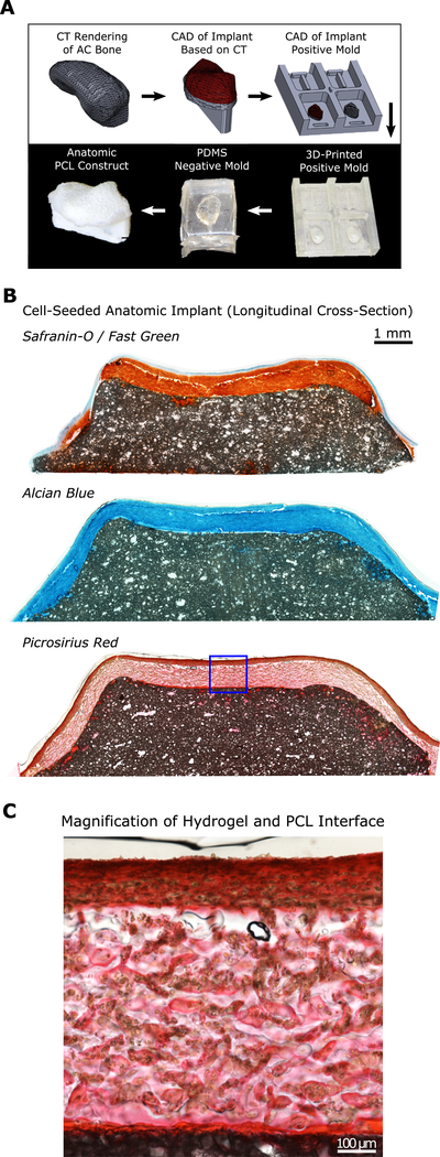
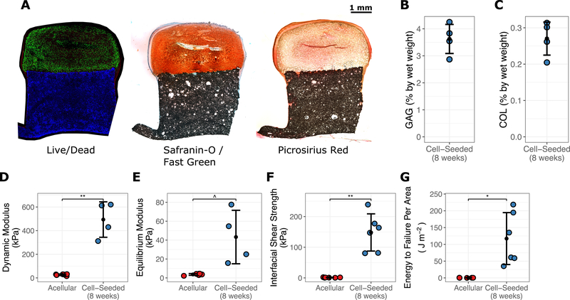
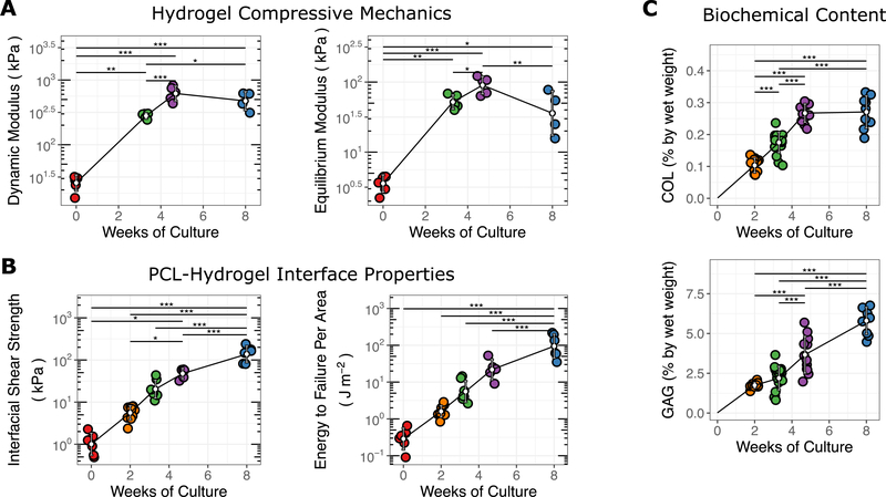
Articular cartilage injury can lead to joint-wide erosion and the early onset of osteoarthritis. To address this, we recently developed a rapid fabrication method to produce patient-specific engineered cartilage tissues to replace an entire articular surface. Here, we extended that work by coupling a mesenchymal stromal cell-laden hydrogel (methacrylated hyaluronic acid) with the porous polycaprolactone (PCL) bone integrating phase and assessed the composition and mechanical performance of these constructs over time. To improve initial construct stability, PCL/hydrogel interface parameters were first optimized by varying PCL pretreatment (with sodium hydroxide before ethanol) before hydrogel infusion. Next, cylindrical osteochondral constructs were formed and cultured in media containing transforming growth factor β3 for up to 8 weeks, with constructs evaluated for viability, histological features, and biochemical content. Mechanical properties were also assessed in axial compression and via an interface shear strength assay. Results showed that the fabrication process was compatible with cell viability, and that construct biochemical content and mechanical properties increased with time. Interestingly, compressive properties peaked at 5 weeks, while interfacial shear properties continued to improve beyond this time point. Finally, these fabrication methods were combined with a custom mold developed from limb-specific computed tomography imaging data to create an anatomic implantable cell-seeded biologic joint surface, which showedmaturation similar to the osteochondral cylinders. Future work will apply these advances in large animal models of critically sized osteochondral defects to study repair and whole joint resurfacing.

摘要

关节软骨损伤可导致关节广泛侵蚀和骨关节炎的早期发作。为了解决这个问题,我们最近开发了一种快速制造方法,以生产个性化的工程软骨组织来替代整个关节表面。在这里,我们通过将富含间充质基质细胞的水凝胶(甲基丙烯酰化透明质酸)与多孔聚己内酯(PCL)骨整合相耦合,扩展了这项工作,并评估了这些构建体随时间的组成和机械性能。为了提高初始构建体的稳定性,首先通过改变 PCL 预处理(在乙醇前用氢氧化钠处理)来优化 PCL/水凝胶界面参数,然后进行水凝胶灌注。接下来,形成圆柱形的骨软骨构建体,并在含有转化生长因子 β3 的培养基中培养长达 8 周,对构建体进行活力、组织学特征和生化含量的评估。还通过轴向压缩和界面剪切强度测定评估了机械性能。结果表明,制造过程与细胞活力兼容,并且构建体的生化含量和机械性能随时间增加。有趣的是,压缩性能在 5 周时达到峰值,而界面剪切性能在该时间点之后继续提高。最后,这些制造方法与从肢体特定的计算机断层扫描成像数据开发的定制模具相结合,创建了可植入细胞的生物关节表面,其成熟度类似于骨软骨圆柱体。未来的工作将在大动物临界尺寸骨软骨缺损模型中应用这些进展,以研究修复和整个关节表面置换。

https://cdn.ncbi.nlm.nih.gov/pmc/blobs/5c34/8222412/c9a02cd1d366/nihms-1657808-f0005.jpg
https://cdn.ncbi.nlm.nih.gov/pmc/blobs/5c34/8222412/5397b4547e4e/nihms-1657808-f0001.jpg
https://cdn.ncbi.nlm.nih.gov/pmc/blobs/5c34/8222412/a3a761ae476d/nihms-1657808-f0002.jpg
https://cdn.ncbi.nlm.nih.gov/pmc/blobs/5c34/8222412/5ef2229b1ea4/nihms-1657808-f0003.jpg
https://cdn.ncbi.nlm.nih.gov/pmc/blobs/5c34/8222412/c86c033a4914/nihms-1657808-f0004.jpg
https://cdn.ncbi.nlm.nih.gov/pmc/blobs/5c34/8222412/c9a02cd1d366/nihms-1657808-f0005.jpg

相似文献

1
Fabrication and maturation of integrated biphasic anatomic mesenchymal stromal cell-laden composite scaffolds for osteochondral repair and joint resurfacing.用于骨软骨修复和关节表面置换的一体化双相解剖间充质基质细胞负载复合支架的制造和成熟。
J Orthop Res. 2021 Nov;39(11):2323-2332. doi: 10.1002/jor.24969. Epub 2021 Jan 14.
2
Tailoring the subchondral bone phase of a multi-layered osteochondral construct to support bone healing and a cartilage analog.定制多层骨软骨构建体的软骨下骨相以支持骨愈合和软骨类似物。
Acta Biomater. 2018 Sep 15;78:351-364. doi: 10.1016/j.actbio.2018.08.009. Epub 2018 Aug 10.
3
Cell-laden hydrogels for osteochondral and cartilage tissue engineering.用于骨软骨和软骨组织工程的载细胞水凝胶。
Acta Biomater. 2017 Jul 15;57:1-25. doi: 10.1016/j.actbio.2017.01.036. Epub 2017 Jan 11.
4
Anatomic Mesenchymal Stem Cell-Based Engineered Cartilage Constructs for Biologic Total Joint Replacement.用于生物全关节置换的基于解剖学间充质干细胞的工程化软骨构建体。
Tissue Eng Part A. 2016 Feb;22(3-4):386-95. doi: 10.1089/ten.tea.2015.0384.
5
Evaluation of an extracellular matrix-derived acellular biphasic scaffold/cell construct in the repair of a large articular high-load-bearing osteochondral defect in a canine model.评价细胞外基质衍生去细胞双相支架/细胞构建体在犬模型中大关节高负荷承重骨软骨缺损修复中的作用。
Chin Med J (Engl). 2011 Dec;124(23):3930-8.
6
3D printing of fibre-reinforced cartilaginous templates for the regeneration of osteochondral defects.用于骨软骨缺损再生的纤维增强软骨模板的3D打印
Acta Biomater. 2020 Sep 1;113:130-143. doi: 10.1016/j.actbio.2020.05.040. Epub 2020 Jun 4.
7
Photo-crosslinked integrated triphasic scaffolds with gradient composition and strength for osteochondral regeneration.用于骨软骨再生的具有梯度组成和强度的光交联集成三相支架。
J Mater Chem B. 2024 Jan 31;12(5):1271-1284. doi: 10.1039/d3tb02031b.
8
Repair and regeneration of osteochondral defects in the articular joints.关节软骨下骨缺损的修复与再生
Biomol Eng. 2007 Nov;24(5):489-95. doi: 10.1016/j.bioeng.2007.07.014. Epub 2007 Aug 7.
9
Fabrication and development of artificial osteochondral constructs based on cancellous bone/hydrogel hybrid scaffold.基于松质骨/水凝胶混合支架的人工骨软骨构建体的制造与开发。
J Mater Sci Mater Med. 2016 Jun;27(6):114. doi: 10.1007/s10856-016-5722-5. Epub 2016 May 14.
10
Investigation of multiphasic 3D-bioplotted scaffolds for site-specific chondrogenic and osteogenic differentiation of human adipose-derived stem cells for osteochondral tissue engineering applications.用于骨软骨组织工程应用的人脂肪来源干细胞的特异性软骨和成骨分化的多相 3D 生物绘制支架的研究。
J Biomed Mater Res B Appl Biomater. 2020 Jul;108(5):2017-2030. doi: 10.1002/jbm.b.34542. Epub 2019 Dec 27.

引用本文的文献

1
Multi-Scale Analysis of Regional Pathology in the Human Trapezium During Osteoarthritis Progression.骨关节炎进展过程中人类大多角骨区域病理学的多尺度分析
J Orthop Res. 2025 Sep;43(9):1581-1591. doi: 10.1002/jor.26112. Epub 2025 Jun 1.
2
Challenges and recent advances in engineering the osteochondral interface.工程化骨软骨界面的挑战与最新进展
Curr Opin Biomed Eng. 2024 Sep;31. doi: 10.1016/j.cobme.2024.100546. Epub 2024 May 18.
3
Enhanced angiogenesis in porous poly(ε-caprolactone) scaffolds fortified with methacrylated hyaluronic acid hydrogel after subcutaneous transplantation.

本文引用的文献

1
Metabolic Labeling to Probe the Spatiotemporal Accumulation of Matrix at the Chondrocyte-Hydrogel Interface.代谢标记法用于探究软骨细胞-水凝胶界面处基质的时空积累情况。
Adv Funct Mater. 2020 Oct 28;30(44). doi: 10.1002/adfm.201909802. Epub 2020 Apr 3.
2
3D Bioprinting of Spatially Heterogeneous Collagen Constructs for Cartilage Tissue Engineering.用于软骨组织工程的空间异质胶原蛋白构建体的3D生物打印
ACS Biomater Sci Eng. 2016 Oct 10;2(10):1800-1805. doi: 10.1021/acsbiomaterials.6b00288. Epub 2016 Aug 4.
3
Magneto-Driven Gradients of Diamagnetic Objects for Engineering Complex Tissues.
皮下移植后,用甲基丙烯酸化透明质酸水凝胶强化的多孔聚(ε-己内酯)支架中血管生成增强。
Biomater Transl. 2024 Mar 28;5(1):59-68. doi: 10.12336/biomatertransl.2024.01.006. eCollection 2024.
4
Osteochondral regenerative engineering: challenges, state-of-the-art and translational perspectives.骨软骨再生工程:挑战、现状与转化前景
Regen Biomater. 2022 Dec 26;10:rbac109. doi: 10.1093/rb/rbac109. eCollection 2023.
5
Systematic review on the application of 3D-bioprinting technology in orthoregeneration: current achievements and open challenges.3D生物打印技术在骨再生中的应用系统综述:当前成果与开放挑战
J Exp Orthop. 2022 Sep 19;9(1):95. doi: 10.1186/s40634-022-00518-3.
6
Gravity-based patterning of osteogenic factors to preserve bone structure after osteochondral injury in a large animal model.基于重力的成骨因子模式在大型动物模型中骨软骨损伤后保持骨结构。
Biofabrication. 2022 Jul 5;14(4). doi: 10.1088/1758-5090/ac79cd.
7
Stem Cell-Laden Hydrogel-Based 3D Bioprinting for Bone and Cartilage Tissue Engineering.用于骨与软骨组织工程的基于载干细胞水凝胶的3D生物打印
Front Bioeng Biotechnol. 2022 May 17;10:865770. doi: 10.3389/fbioe.2022.865770. eCollection 2022.
8
Directed Regeneration of Osteochondral Tissue by Hierarchical Assembly of Spatially Organized Composite Spheroids.通过空间组织的复合球体的层次组装指导骨软骨组织的再生。
Adv Sci (Weinh). 2022 Jan;9(3):e2103525. doi: 10.1002/advs.202103525. Epub 2021 Nov 21.
9
The porcine accessory carpal bone as a model for biologic joint replacement for trapeziometacarpal osteoarthritis.猪腕侧副骨作为掌指关节骨关节炎生物型关节置换的模型。
Acta Biomater. 2021 Jul 15;129:159-168. doi: 10.1016/j.actbio.2021.05.011. Epub 2021 May 19.
磁驱动抗磁物体各向异性用于工程复杂组织。
Adv Mater. 2020 Dec;32(48):e2005030. doi: 10.1002/adma.202005030. Epub 2020 Oct 19.
4
Optimized Media Volumes Enable Homogeneous Growth of Mesenchymal Stem Cell-Based Engineered Cartilage Constructs.优化的培养基体积可实现基于间充质干细胞的工程化软骨构建体的均匀生长。
Tissue Eng Part A. 2021 Feb;27(3-4):214-222. doi: 10.1089/ten.TEA.2020.0123. Epub 2020 Nov 2.
5
Autologous Chondrocyte Implantation as Treatment for Unsalvageable Osteochondritis Dissecans: 10- to 25-Year Follow-up.自体软骨细胞移植治疗不可挽救的剥脱性骨软骨炎:10 至 25 年随访结果。
Am J Sports Med. 2020 Apr;48(5):1134-1140. doi: 10.1177/0363546520908588. Epub 2020 Mar 17.
6
Emerging therapies for cartilage regeneration in currently excluded 'red knee' populations.针对目前被排除在外的“红色膝关节”人群的软骨再生新兴疗法。
NPJ Regen Med. 2019 May 30;4:12. doi: 10.1038/s41536-019-0074-7. eCollection 2019.
7
A soft 3D polyacrylate hydrogel recapitulates the cartilage niche and allows growth-factor free tissue engineering of human articular cartilage.一种柔软的 3D 聚丙烯酸酯水凝胶再现了软骨小生境,并允许在无生长因子的情况下进行人关节软骨的组织工程。
Acta Biomater. 2019 May;90:146-156. doi: 10.1016/j.actbio.2019.03.040. Epub 2019 Mar 22.
8
Fabrication and mechanical characterization of 3D printed vertical uniform and gradient scaffolds for bone and osteochondral tissue engineering.用于骨和骨软骨组织工程的 3D 打印垂直均匀和梯度支架的制造和机械特性。
Acta Biomater. 2019 May;90:37-48. doi: 10.1016/j.actbio.2019.03.041. Epub 2019 Mar 21.
9
Biofabrication for osteochondral tissue regeneration: bioink printability requirements.用于骨软骨组织再生的生物制造:生物墨水可打印性要求。
J Mater Sci Mater Med. 2019 Jan 28;30(2):20. doi: 10.1007/s10856-019-6218-x.
10
Advances in Porous Scaffold Design for Bone and Cartilage Tissue Engineering and Regeneration.多孔支架设计在骨和软骨组织工程和再生中的研究进展。
Tissue Eng Part B Rev. 2019 Feb;25(1):14-29. doi: 10.1089/ten.TEB.2018.0119. Epub 2018 Sep 20.