Falk Martin, Hausmann Michael
Institute of Biophysics, The Czech Academy of Sciences, 612 65 Brno, Czech Republic.
Kirchhoff Institute for Physics, Heidelberg University, 69120 Heidelberg, Germany.
Cancers (Basel). 2020 Dec 23;13(1):18. doi: 10.3390/cancers13010018.
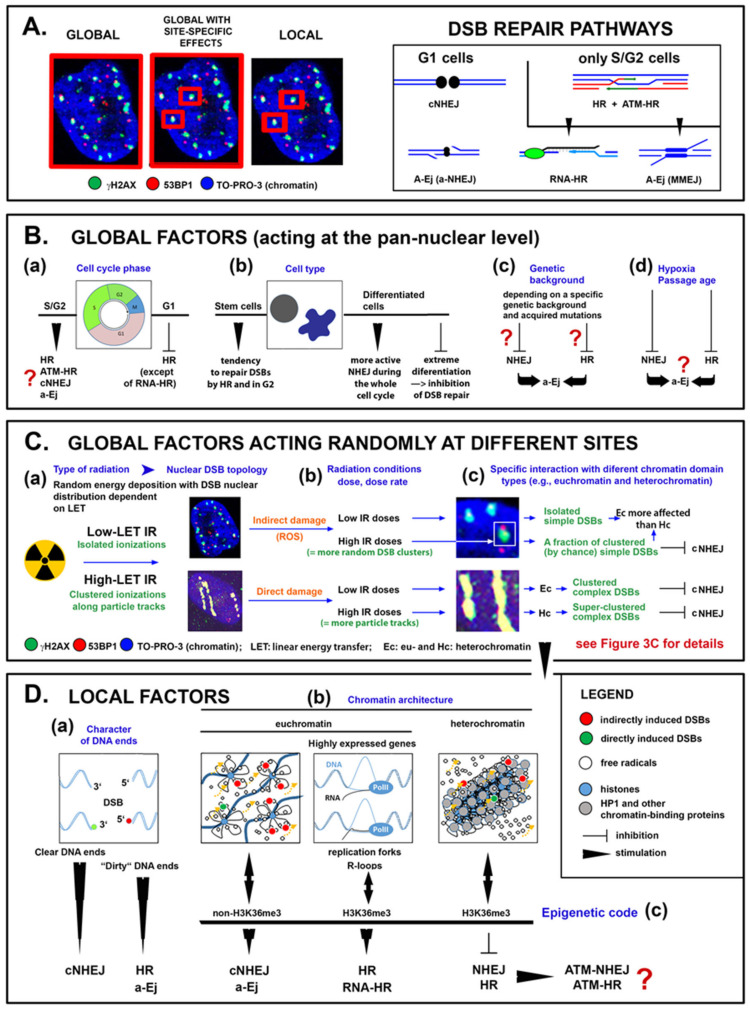
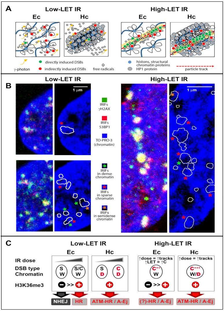
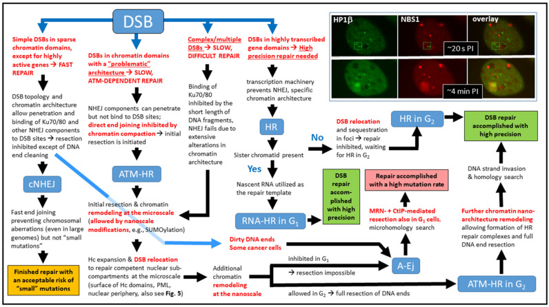
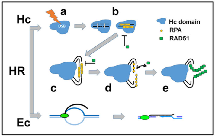
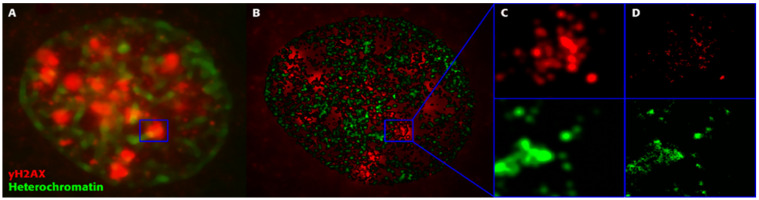
DNA double-strand breaks (DSBs) have been recognized as the most serious lesions in irradiated cells. While several biochemical pathways capable of repairing these lesions have been identified, the mechanisms by which cells select a specific pathway for activation at a given DSB site remain poorly understood. Our knowledge of DSB induction and repair has increased dramatically since the discovery of ionizing radiation-induced foci (IRIFs), initiating the possibility of spatiotemporally monitoring the assembly and disassembly of repair complexes in single cells. IRIF exploration revealed that all post-irradiation processes-DSB formation, repair and misrepair-are strongly dependent on the characteristics of DSB damage and the microarchitecture of the whole affected chromatin domain in addition to the cell status. The microscale features of IRIFs, such as their morphology, mobility, spatiotemporal distribution, and persistence kinetics, have been linked to repair mechanisms. However, the influence of various biochemical and structural factors and their specific combinations on IRIF architecture remains unknown, as does the hierarchy of these factors in the decision-making process for a particular repair mechanism at each individual DSB site. New insights into the relationship between the physical properties of the incident radiation, chromatin architecture, IRIF architecture, and DSB repair mechanisms and repair efficiency are expected from recent developments in optical superresolution microscopy (nanoscopy) techniques that have shifted our ability to analyze chromatin and IRIF architectures towards the nanoscale. In the present review, we discuss this relationship, attempt to correlate still rather isolated nanoscale studies with already better-understood aspects of DSB repair at the microscale, and consider whether newly emerging "correlated multiscale structuromics" can revolutionarily enhance our knowledge in this field.
DNA双链断裂(DSB)已被公认为是受辐照细胞中最严重的损伤。虽然已经确定了几种能够修复这些损伤的生化途径,但细胞在给定DSB位点选择特定途径进行激活的机制仍知之甚少。自从发现电离辐射诱导灶(IRIF)以来,我们对DSB诱导和修复的认识有了显著提高,这开启了在单细胞中对修复复合物的组装和拆卸进行时空监测的可能性。对IRIF的探索表明,除细胞状态外,所有辐照后过程——DSB形成、修复和错配修复——都强烈依赖于DSB损伤的特征以及整个受影响染色质结构域的微观结构。IRIF的微观特征,如它们的形态、移动性、时空分布和持续动力学,已与修复机制相关联。然而,各种生化和结构因素及其特定组合对IRIF结构的影响仍然未知,这些因素在每个单独DSB位点特定修复机制的决策过程中的层级关系也同样未知。光学超分辨率显微镜(纳米显微镜)技术的最新进展有望为入射辐射的物理性质、染色质结构、IRIF结构以及DSB修复机制和修复效率之间的关系带来新的见解,这些进展已将我们分析染色质和IRIF结构的能力提升至纳米尺度。在本综述中,我们讨论了这种关系,试图将仍然较为孤立的纳米尺度研究与微观尺度上已被更好理解的DSB修复方面相关联,并考虑新出现的“相关多尺度结构组学”是否能革命性地增强我们在该领域的知识。